

While sinus arrhythmia is often reflexively grouped under normal physiological variants, clinically, it presents in three distinct forms driven by different mechanisms.
Here is a breakdown of the three primary types of sinus arrhythmia.
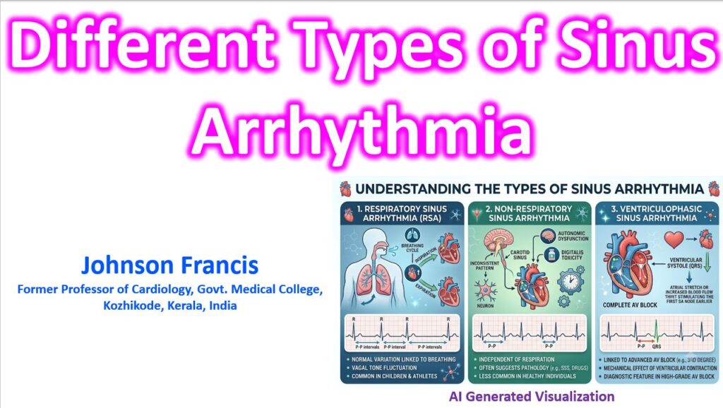
1. Respiratory Sinus Arrhythmia (RSA)
This is the most common and widely recognized form, representing a normal, physiological phenomenon heavily influenced by autonomic regulation.
- Mechanism: RSA is driven by variations in vagal tone during the respiratory cycle. During inspiration, reflex inhibition of vagal tone occurs, leading to an increased heart rate (shortened P-P intervals). During expiration, vagal tone is restored, slowing the heart rate (lengthened P-P intervals).
- ECG Findings: Gradual lengthening and shortening of the P-P intervals that sync exactly with the patient’s respiratory rate. All P waves have a normal sinus morphology.
- Clinical Context: It is a marker of robust autonomic tone and is most pronounced in children, young adults, and athletes. It typically diminishes with age or with conditions that cause autonomic neuropathy (like diabetes).
Key insight: The presence of a robust RSA can actually be a useful clinical indicator of healthy parasympathetic function, while its sudden absence in a younger patient might warrant a closer look.
2. Non-Respiratory Sinus Arrhythmia
Unlike RSA, this form is completely independent of the respiratory cycle. The variations in the P-P intervals are erratic or cyclical but do not correlate with breathing phases.
- Mechanism: The exact mechanism is variable. It often reflects a depressed sinus node automatism or altered parasympathetic/sympathetic balance independent of stretch receptors in the lungs.
- Clinical Context: While it can occasionally be a normal variant in older individuals or during periods of increased vagal tone (like sleep), it frequently carries pathological significance.
- Common Etiologies:
- Digitalis toxicity
- Acute inferior wall myocardial infarction (due to ischemia of the SA node or heightened vagal reflexes)
- Increased intracranial pressure (often manifesting as part of the Cushing reflex)
- Early stages of Sick Sinus Syndrome (often preceding sinus pauses or tachy-brady syndrome)
3. Ventriculophasic Sinus Arrhythmia
This is a specific, mechanically-driven type of sinus arrhythmia that only occurs in the setting of advanced atrioventricular (AV) block, most notably complete (third-degree) heart block.
- ECG Findings: The defining characteristic is that the P-P interval containing a QRS complex is measurably shorter than the P-P interval that does not enclose a QRS complex.
- Mechanism: It is thought to be driven by the mechanical effects of ventricular systole. The current leading theories suggest that ventricular contraction either increases local blood flow to the SA node (briefly increasing its firing rate) or stretches the right atrium, which mechanically stimulates the SA node to fire slightly earlier via a local reflex arc.