
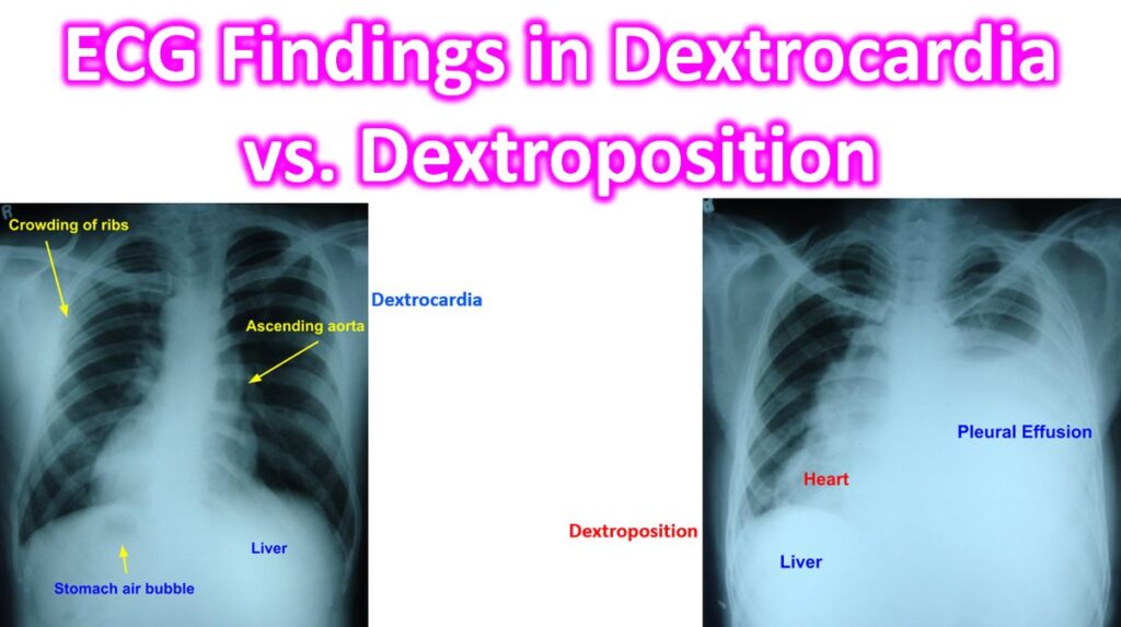

Distinguishing between Dextrocardia (a congenital positional anomaly where the heart is mirrored) and Dextroposition (where the heart is displaced to the right by external factors like lung collapse or a tumor) is a classic clinical challenge. While both result in the heart being in the right chest, their ECG signatures are fundamentally different.
1. Dextrocardia (Situs Inversus)
In true dextrocardia, the heart’s electrical axis is mirrored. The most striking feature is that the ECG looks “upside down” in the limb leads and “backward” in the precordial leads.
- Lead I: Global inversion. You will see an inverted P wave, a negative QRS complex, and an inverted T wave. (In a normal heart, this usually suggests the limb leads were accidentally swapped).
- Leads aVR and aVL: These effectively “swap” roles. The P and QRS will be positive in aVR (which is normally negative) and negative in aVL.
- Precordial Progression (V1–V6): There is a reverse R-wave progression. Instead of the R-wave getting taller from V1 to V6, it starts small in V1 and disappears or stays flat toward V6 because the heart is away from the left-sided electrodes.
Clinical Tip: To confirm dextrocardia and get a “normal” looking reading, you must perform a Right-Sided ECG by placing the precordial leads (V3R–V6R) on the right side of the chest and swapping the left and right arm electrodes.
2. Dextroposition
In dextroposition, the heart is structurally “normal” (not mirrored) but has been physically pushed or pulled into the right hemithorax.
- Axis: The electrical axis usually remains relatively normal or shows a slight shift, but it does not show the global Lead I inversion seen in dextrocardia.
- P Waves: The P wave in Lead I remains upright/positive because the SA node is still in its anatomical position relative to the left/right orientation.
- Precordial Progression: Like dextrocardia, you will see low voltage or poor R-wave progression in the left-sided leads (V4–V6) because the heart is physically distant from those electrodes. However, V1 and V2 might show unexpectedly high voltage as they are now closer to the bulk of the displaced heart.
Summary Comparison Table
| Feature | Dextrocardia (Situs Inversus) | Dextroposition |
| Lead I P wave | Inverted (Negative) | Upright (Positive) |
| Lead I QRS | Negative / Inverted | Usually Positive |
| aVR / aVL | Roles Swapped (aVR is positive) | Normal orientation |
| Cause | Congenital mirroring | Displacement (e.g., Pneumothorax, Scoliosis) |
| R-wave Progression | Progressively decreases V1 – V6 | Low voltage in V4–V6; P wave remains normal |