
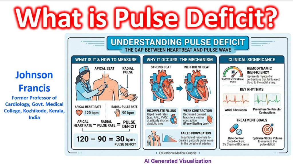

A pulse deficit is the clinical finding where the heart rate (counted by listening to the apical beat with a stethoscope) is higher than the peripheral pulse rate (counted by palpating the radial artery) when measured simultaneously.
Mathematically, it is:
Pulse Deficit = Apical Heart Rate – Radial Pulse Rate
This is most frequently seen in patients with Atrial Fibrillation (AFib), though it also occurs with frequent premature ventricular contractions (PVCs) or premature atrial contractions (PACs).
Hemodynamic Mechanism
A pulse deficit represents mechanically wasted myocardial work. It happens when a cardiac cycle initiates electrical depolarization (seen on an ECG) and a subsequent ventricular contraction (auscultated as a heartbeat), but that contraction fails to generate a palpable peripheral pulse wave. Here is the step-by-step breakdown of why it occurs:
- Shortened Diastole: In rhythms like AFib with a rapid ventricular response or early ectopic beats, the RR interval is highly variable. When an RR interval is exceptionally short, diastolic filling time is drastically reduced.
- Inadequate Preload: Because the left ventricle doesn’t have enough time to fill, the end-diastolic volume (preload) is extremely low.
- Failed Ejection: By the Frank-Starling law, the resulting ventricular contraction is weak. The left ventricle may contract enough to produce a first heart sound (S1), but the stroke volume is insufficient to generate enough pressure to open the aortic valve—or, if it does open, the stroke volume is too small to propagate a palpable pressure wave all the way to the radial artery.
Clinical Significance
In clinical practice, a large pulse deficit indicates significant hemodynamic inefficiency.
- In Atrial Fibrillation: It reflects the degree of chaotic, rapid conduction and loss of the “atrial kick.” As you control the ventricular rate (e.g., with beta-blockers or calcium channel blockers), diastolic filling time improves, stroke volume normalizes, and the pulse deficit typically shrinks or disappears.
- In Ectopy: Frequent PVCs often result in a “dropped” radial pulse for the ectopic beat, followed by a stronger-than-normal compensatory beat due to prolonged filling time.