Apical five chamber view in Tetralogy of Fallot – echocardiogram
Apical five chamber view in Tetralogy of Fallot – echocardiogram
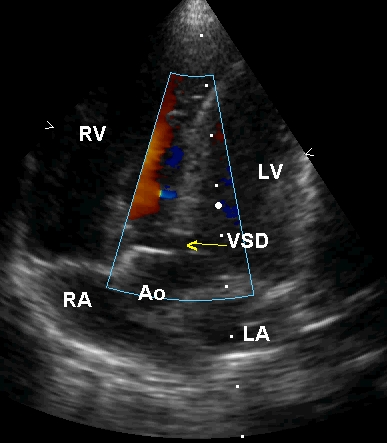
Apical five chamber view in Tetralogy of Fallot demonstrating the sub aortic ventricular septal defect (VSD) with aortic over-ride. 50% of the aorta (Ao) is committed to the left ventricle (LV) while the remaining half is committed to the right ventricle (RV). LA: left atrium; RA: right atrium. The VSD in Tetralogy of Fallot is a mal-allignment VSD which results from the mal-allignment of the ventricular septum with respect to the aortico-pulmonary septum during embryonic development. The shift of the aortico pulmonary septum towards the pulmonary side produces both the ventricular septal defect and the narrowing of the right ventricular outflow tract. This theory is sometimes termed the Monology of Fallot meaning that all the four defects in Tetralogy of Fallot (ventricular septal defect, over-riding aorta, right ventricular outflow tract obstruction and hypertrophy of the right ventricle) are in fact due to one defect in the embryonic development.
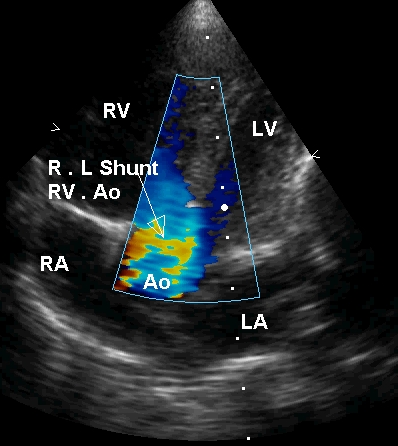
Apical five chamber view in Tetralogy of Fallot with colour flow mapping (Colour Doppler imaging) in systole with right to left shunt across the VSD. Blue stream moving from right ventricle across the VSD to the aorta is clearly visualised in this frame. There is also a blue stream from the left ventricle to the aorta.
Related Posts


About The Author
Johnson Francis
Former Professor of Cardiology, Calicut Govt. Medical Kozhikode, Kerala, India. Editor-in-Chief, BMH Medical Journal