


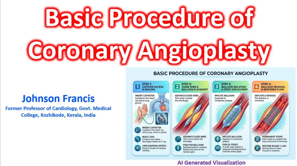

Here is a step-by-step breakdown of a Percutaneous Coronary Intervention (PCI) or Coronary Angioplasty. It is used to remove major blocks in the coronary arteries, mostly for symptom relief and a life saving procedure in situations like a heart attack.
1.Arterial Access:Usually via the radial artery.
After administering local anesthesia and mild conscious sedation, a small puncture is made. This is most commonly done in the radial artery in the wrist, though the femoral artery in the groin is a traditional alternative. A small sheath is inserted to serve as the working portal for the entire procedure.
2.Catheter Navigation and Angiography:Mapping the blockages.
A long, flexible tube (diagnostic catheter) is threaded through the arterial system up to the ostium of the coronary arteries. Contrast dye is injected under continuous X-ray (fluoroscopy) to create a roadmap of the heart’s vessels, pinpointing the exact location and severity of any stenotic plaques.
3.Crossing the Lesion:
Once the blockage is identified, the diagnostic catheter is swapped for a guiding catheter. A thin, highly steerable guidewire (typically 0.014 inches in diameter) is carefully navigated through the catheter and advanced across the narrowed segment of the artery. This wire acts as a monorail for the therapeutic equipment that follows.
4.Balloon Angioplasty:Pre-dilation.
A deflated balloon catheter is tracked over the guidewire directly into the center of the blockage. The balloon is then inflated at high atmospheric pressure for a few seconds. This physically compresses the calcified or fatty atherosclerotic plaque against the artery walls, creating a wider channel for blood flow.
5.Stent Deployment:Providing structural support.
To prevent the artery from recoiling or re-narrowing, a stent (a tiny, expandable metallic wire mesh tube) is delivered to the site. Modern stents are usually drug-eluting, coated with medication to prevent tissue overgrowth. The stent is mounted on another balloon; when inflated, the stent expands and locks into place against the arterial wall, acting as a permanent scaffold.
6.Final Angiogram and Closure:
The balloon and guidewire are removed, leaving the stent permanently embedded in the vessel wall. A final contrast injection confirms that normal blood flow is fully restored and the stent is properly expanded. The sheath is then removed, and a compression band or closure device is applied to the access site to achieve complete hemostasis.