


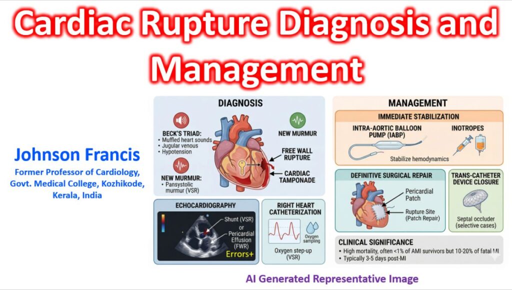

Cardiac rupture is a catastrophic complication typically occurring after a myocardial infarction (MI). It often involves a full-thickness tear of the myocardium, leading to the escape of blood into the pericardial space or adjacent cardiac chambers. Recent medical literature emphasizes that while the incidence of cardiac rupture has decreased due to advanced reperfusion strategies, its mortality remains high, often exceeding 90%.
Classification by Anatomical Site
Cardiac rupture is categorized based on which part of the heart is affected:
- Free Wall Rupture (FWR): The most common and lethal form. Blood leaks into the pericardial sac, causing rapid cardiac tamponade.
- Ventricular Septal Rupture (VSR): A tear in the wall between the left and right ventricles, creating an acquired left-to-right shunt. This leads to acute right heart failure and pulmonary edema.
- Papillary Muscle Rupture: The tearing of the muscles supporting the mitral valve, resulting in acute, severe mitral regurgitation and cardiogenic shock.
Echocardiographic Classification of Papillary Muscle Rupture (PMR)
Partial-incomplete: Partially ruptured papillary muscle remains adherent to the ventricular wall and exhibits chaotic movement.
Partial-complete: Ruptured portion of the papillary muscle is freely mobile within the left ventricle but does not protrude into the left atrium during systole.
Subtotal/total: Heads or the entire trunk of the papillary muscle protrudes into the left atrium during systole, pulling the chordae tendineae and mitral leaflets along.
Clinical Presentation & Risk Factors
While the timing can vary, these events most frequently occur 3 to 5 days post-MI, when the necrotic tissue is softest (yellow softening) but before significant collagen scarring has developed.
Common Signs
- Sudden Hemodynamic Collapse: Rapid onset of hypotension and shock.
- Beck’s Triad (for FWR): Jugular venous distention, muffled heart sounds, and hypotension.
- New Harsh Murmur: A “pansystolic” murmur is characteristic of VSR or papillary muscle rupture.
Patient Profile at Higher Risk
- First-time MI (lack of collateral circulation).
- Anterior wall infarction.
- Advanced age and female gender.
- Hypertension during the acute phase of MI.
- Delayed reperfusion therapy.
Diagnosis and Management
| Tool | Findings |
| Echocardiography | The gold standard. Shows pericardial effusion (FWR), a shunt (VSR), or a flail mitral leaflet. |
| Right Heart Cath | Shows an “oxygen step-up” in the right ventricle in the case of a VSR. |
| ECG | May show persistent ST-elevation or “electromechanical dissociation” (Pulseless Electrical Activity). |
Emergency Management
- Stabilization: Use of inotropes or an Intra-Aortic Balloon Pump (IABP) to reduce afterload and improve coronary perfusion. This is one of the few situations in which IABP still has a role.
- Pericardiocentesis: If tamponade is present, though this is often a temporary measure for free wall ruptures.
- Surgery: Definitive treatment requires urgent surgical repair (e.g., infarct excision and patching). Some centers use transcatheter device closure for selected cases of ventricular septal rupture. Both options are technically difficult because of the friability of border tissue.