


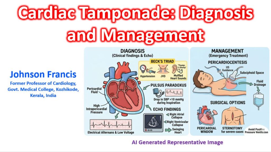

Cardiac tamponade is a life-threatening medical emergency where fluid accumulates in the pericardial sac, increasing intrapericardial pressure to the point that it exceeds intracardiac pressure, leading to impaired diastolic filling and reduced cardiac output.
Diagnosis
The diagnosis is primarily clinical, supported by bedside imaging. Because it is a dynamic process, a high index of suspicion is required in patients with known risk factors (e.g., malignancy, recent cardiac surgery, or trauma).
Clinical Findings
- Beck’s Triad: The classic (though not always present) trio of:
- Hypotension (due to decreased stroke volume).
- Jugular Venous Distension (due to impaired venous return).
- Muffled heart sounds (due to the insulating effect of the fluid).
- Pulsus Paradoxus: A drop in systolic blood pressure of >10 mmHg during inspiration. This is a hallmark sign but can be absent in patients with ASD or severe aortic regurgitation.
- Tachycardia: A compensatory mechanism to maintain cardiac output.
Diagnostic Investigations
- Echocardiography (Gold Standard): * Right Atrial (RA) collapse: Occurs in late diastole (earliest sign).
- Right Ventricular (RV) collapse: Occurs in early diastole (highly specific).
- Plethoric IVC: A dilated Inferior Vena Cava with <50% inspiratory collapse (indicates high CVP).
- Swinging Heart: Large effusions may cause the heart to oscillate within the sac.
- ECG: May show Electrical Alternans (varying QRS amplitude) and low voltage QRS complexes.
- Chest X-ray: May show a “water bottle” heart silhouette, though this is only visible once the effusion is large (>200–250 mL).
Management
The definitive treatment for cardiac tamponade is the removal of pericardial fluid to relieve the pressure.
Immediate Stabilization
- Volume Expansion: Intravenous fluid boluses (Normal Saline) can temporarily increase preload and help maintain cardiac output while preparing for drainage.
- Avoid Positive Pressure Ventilation: Manual or mechanical ventilation can further decrease venous return and precipitate circulatory collapse. If intubation is necessary, it must be done with extreme caution.
Definitive Procedures
- Pericardiocentesis: * Usually performed under ultrasound guidance (Subxiphoid or Apical approach).
- A catheter is often left in place for continuous drainage until the output is minimal.
- Pericardial Window: * A surgical procedure where a small piece of the pericardium is removed.
- Indicated for recurrent effusions, clotted blood (hemopericardium), or when a biopsy is needed.
- Emergency Sternotomy/Thoracotomy: Reserved for traumatic tamponade or post-surgical cases where rapid access is required to control hemorrhage.
“Low-Pressure” Tamponade
It is worth noting that in patients who are severely dehydrated or hypovolemic, the classic signs (like JVD) may be absent because the intracardiac pressures are low. This is “low-pressure tamponade,” where even a small amount of fluid can cause collapse because the heart isn’t “full” enough to push back against the effusion.