


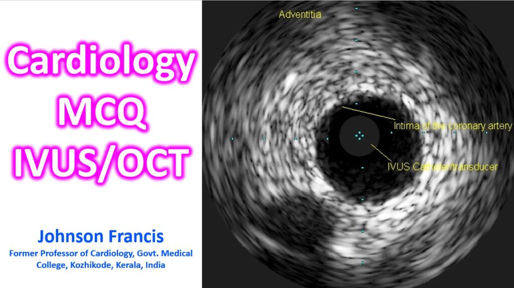

Question 1: Fundamental Principles
Which of the following physical characteristics primarily distinguishes OCT from IVUS imaging?
A) OCT uses high-frequency sound waves, whereas IVUS utilizes near-infrared light.
B) OCT relies on near-infrared light and requires blood clearance, while IVUS uses ultrasound and does not require blood clearance.
C) OCT provides deeper tissue penetration, whereas IVUS provides 10 times higher axial resolution.
D) Both modalities rely on the same acoustic principles but different catheter sizes.
Correct Answer: B
Explanation: OCT uses near-infrared light, which has a shorter wavelength than red blood cells, causing signal backscattering. Thus, it requires temporary blood clearance with contrast or dextran to visualize the vessel wall. IVUS uses high-frequency ultrasound, which does not get blocked by red blood cells.
Question 2: Resolution and Penetration
If a cardiologist needs to accurately measure the thickness of a fibrous cap to identify a vulnerable plaque, which modality is preferred and why?
A) IVUS, because it has deeper tissue penetration of 5 to 6 mm.
B) OCT, because it has an axial resolution of 10-20 μ m versus IVUS’s 100-150 μ m.
C) OCT, because it does not require blood clearance.
D) IVUS, because it provides real-time 3D reconstruction without artifacts.
Correct Answer: B
Explanation: OCT offers approximately 10 times higher axial resolution than IVUS, making it the superior choice for evaluating thin-cap fibroatheromas and fine surface structures.
Question 3: Clinical Strengths
When evaluating a heavily calcified coronary lesion, IVUS is generally preferred over OCT because:
A) IVUS ultrasound waves penetrate through thick calcium, whereas OCT light is blocked.
B) OCT provides better stent strut visualization.
C) IVUS can automatically measure lumen contours with higher reproducibility.
D) IVUS uses light signals that yield superior plaque characterization.
Correct Answer: A
Explanation: While OCT offers superior surface imaging, ultrasound waves from IVUS can penetrate deeper through heavily calcified lesions to evaluate the full depth and arc of the calcium, making it better for assessing deep vessel structures.
Question 4: Clinical Applications
Which of the following is considered an advantage of OCT compared to IVUS?
A) Superior ability to image the External Elastic Membrane (EEM) in large vessels.
B) Deep tissue penetration to evaluate large lipid cores.
C) High-definition imaging of stent malapposition, tissue prolapse, and edge dissection.
D) The procedure can be performed without a guide catheter.
Correct Answer: C
Explanation: Due to its ultra-high resolution, OCT is highly accurate at detecting early post-stent complications, such as incomplete stent apposition, edge dissections, and intraluminal thrombi.