Cardiology X-Ray quiz-Prosthetic valve
Cardiology X-Ray quiz-Prosthetic valve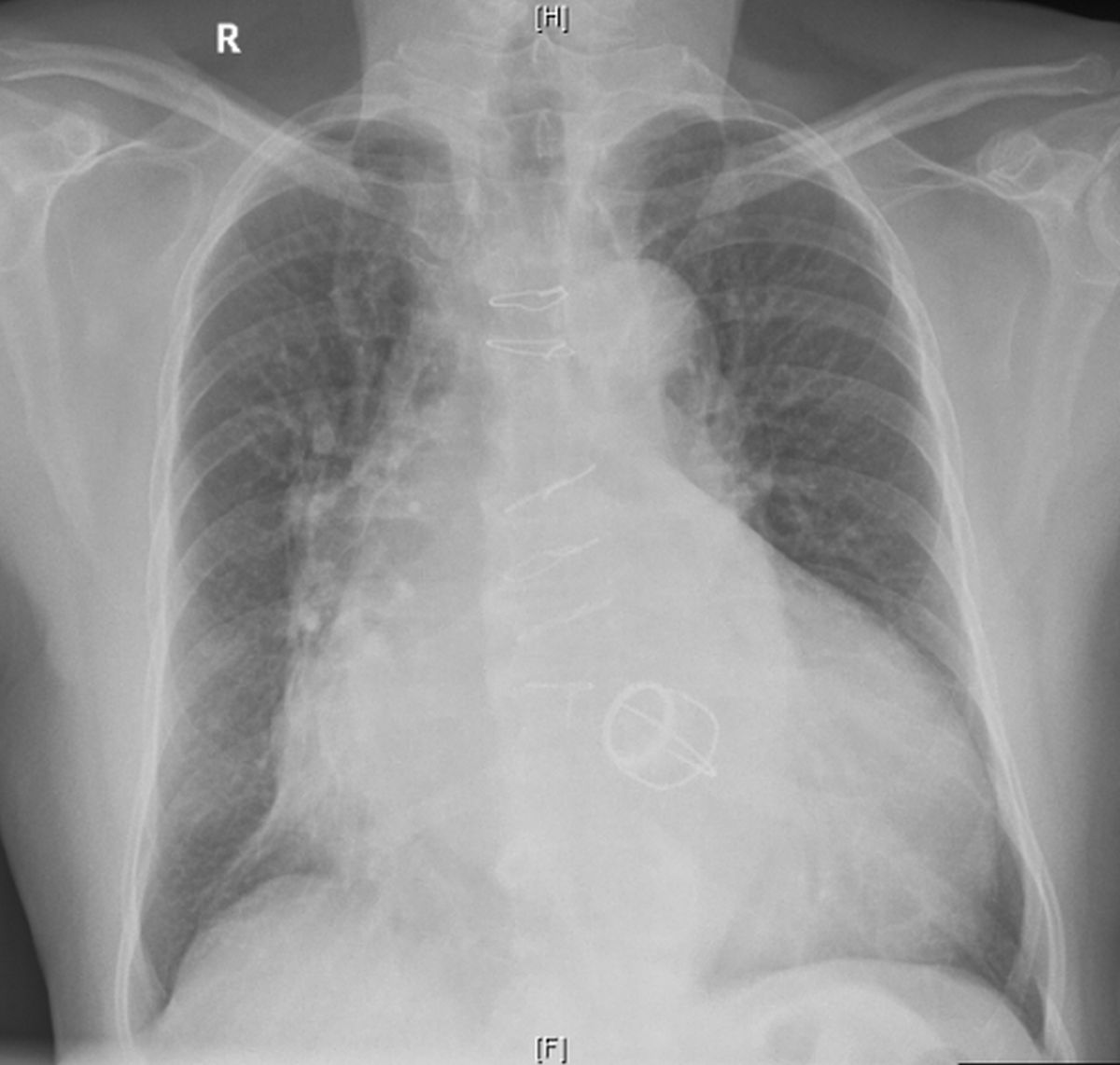
What type of prosthetic valve is seen on this chest X-ray?
Discussion
It is a caged ball prosthesis with four struts facing downwards and to the apex, suggestive of mitral location. The valve is below the line joining the pulmonary bay and right cardiophrenic angle. Aortic prosthesis is situated above this line. Prominent ascending aorta and aortic knuckle as well as sternotomy suture wires are visible. Right atrium is enlarged, with a shadow within shadow indicating co-existing left atrial enlargement. Both these can be a sequelae of previous mitral valve disease. Left atrial appendage is not prominent on the left cardiac border, possibly because it has been removed during a previous closed surgical mitral valvotomy.
Significant cardiomegaly with enhanced cardiothoracic ratio is also visible. This in addition to findings of double atrial enlargement could mean either other valvular lesions which have progressed later or paraprosthetic mitral regurgitation. Some also develop significant left ventricular dysfunction in the long run if the ventricular myocardium had undergone fibrosis due to long term load of the previous valvular lesion, which may not recover fully after surgery. A prominent descending aortic shadow is also seen behind the cardiac silhouette. Aortic dilatation along with cardiomegaly and left atrial enlargement may indicate a pre-existing aortic regurgitation which has now become severe. Atherosclerotic changes in aorta as age advances, is also a possibility.
Though Starr–Edwards prosthetic valves have now gone out of production, some remarkable long term performance has been reported. A patient who was undergoing mitral valve replacement was found to have an aortic Starr–Edwards prosthetic valve which was implanted 51 years back and still functioning well and left in situ [1]. Though such instances are there, SEP has now been supplanted by lower profile bileaflet valves with better hemodynamic profile, lesser risk of thromboembolic complications and longer durability, which only time will tell.
Reference
- Amrane M, Soulat G, Carpentier A, Jouan J. Starr-Edwards aortic valve: 50+ years and still going strong: a case report. Eur Heart J Case Rep. 2017 Dec 20;1(2):ytx014.
Related Posts

About The Author
Johnson Francis
Former Professor of Cardiology, Calicut Govt. Medical Kozhikode, Kerala, India. Editor-in-Chief, BMH Medical Journal
A
B. Starr Edwards prosthetic aortic valve.
Sir, there is prominent cardiomegaly. Left ventricle and ascending aorta also seem to be enlarged. Possibility of aortic valve replacement for aortic stenosis. Starr Edwards valve can be identified by the radioopaque “cage” of the caged ball valve.
Dr.Sanjay Natarajan. MBBS.
It is a caged ball prosthesis with four struts facing downwards and to the apex, suggestive of mitral location. The valve is below the line joining the pulmonary bay and right cardiophrenic angle. Aortic prosthesis is situated above this line. Prominent ascending aorta and aortic knuckle as well as sternotomy suture wires are visible. Right atrium is enlarged, with a shadow within shadow indicating co-existing left atrial enlargement. Both these can be a sequel of previous mitral valve disease. Left atrial appendage is not prominent on the left cardiac border, possibly because it has been removed during a previous closed surgical mitral valvotomy.
Correct answer