Cardiology X-ray Quiz
Cardiology X-ray Quiz
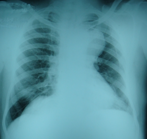
What are the findings and diagnostic possibilities?
Rounded shadow in the left upper zone on this X-ray chest PA view is due to an aortic arch aneurysm. Another possibility is an aneurysm of the ductus arteriosus, which can also rarely occur after surgical closure of patent ductus arteriosus. Lymph nodes in the region as in lymphoma can also produce a similar appearance. An aneurysm in this location can produce hoarseness of voice by left vocal cord palsy as a result of compression of the left recurrent laryngeal nerve. Thymic mass is another differential diagnosis for a lesion in this location. An aneurysmally dilated pulmonary artery would be located slightly lower down.
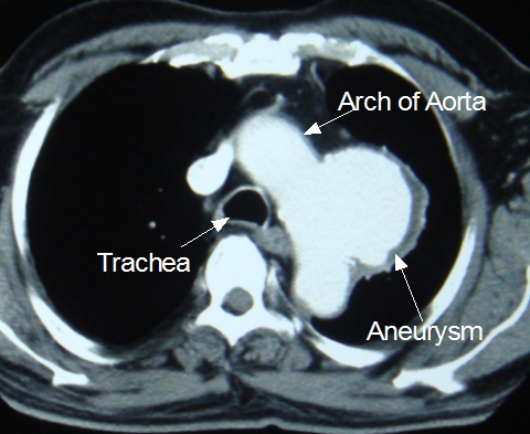
CT scan of thorax showing aortic arch aneurysm. The single tracheal air column identifies the level above the tracheal bifurcation (below the bifurcation, two air columns of the right and left bronchi would be seen). The negative shadow outside the contrast filled region is likely to be an adherent thrombus within the aneurysm. CT appearance is suggestive of a saccular aortic aneurysm with a wide mouth, arising from the aortic arch.
Related Posts


About The Author
Johnson Francis
Former Professor of Cardiology, Calicut Govt. Medical Kozhikode, Kerala, India. Editor-in-Chief, BMH Medical Journal