


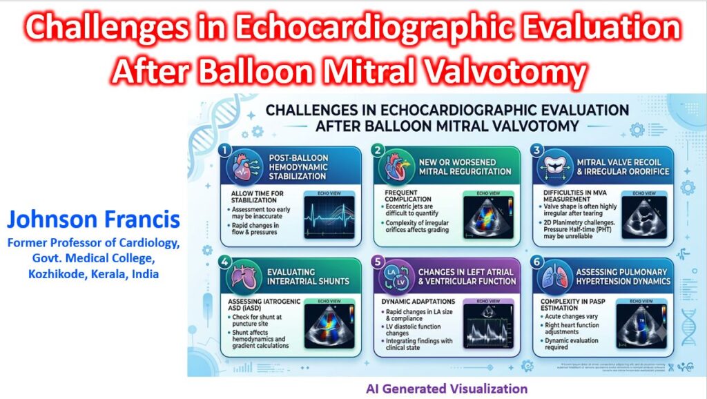

Evaluating a patient immediately after Balloon Mitral Valvotomy (BMV/PTMC) is one of the more hemodynamically complex scenarios in the echo lab. Because the procedure abruptly alters valve geometry, chamber compliance, and interatrial shunting, the standard resting measurements we rely on often contradict each other.
1. The Disconnect in Mitral Valve Area (MVA)
Immediately post-BMV, the reliable triad of planimetry, Pressure Half-Time (PHT), and the continuity equation frequently give highly discordant values.
- Pressure Half-Time (PHT) Inaccuracy: This is the most significant pitfall. PHT relies on a stable relationship between left atrial (LA) and left ventricular (LV) compliance. BMV causes an acute, sudden drop in LA pressure and a rapid increase in early diastolic transmitral flow, temporarily decoupling this relationship. Consequently, PHT often significantly overestimates the MVA in the first 48–72 hours.
- Irregular Orifice in Planimetry: While 2D planimetry remains the anatomical gold standard, successful commissural splitting leaves a complex, irregular, or “tear-drop” shaped orifice. Gaining a true short-axis cross-section exactly at the tips of the newly torn leaflets is technically demanding.
- Continuity Equation Confounders: If the procedure induces Mitral Regurgitation (MR) transmitral stroke volume no longer equals aortic stroke volume, rendering the continuity equation mathematically invalid.
2. Quantifying Post-Procedural Mitral Regurgitation
BMV inevitably alters the coaptation line. While mild MR is a common and acceptable trade-off for a larger valve area, acute, severe MR requires immediate intervention.
- Eccentric Jets: New MR post-BMV is rarely a clean, central jet. Commissural tearing or subvalvular damage creates highly eccentric jets that hug the LA wall. Relying on color flow jet area alone will drastically underestimate the severity.
- Multiple Orifices: You will frequently encounter a pre-existing central regurgitant jet combined with a new, distinct commissural jet. Proximal Isovelocity Surface Area (PISA) and Vena Contracta (VC) become highly flawed when trying to quantify multiple, non-circular regurgitant orifices simultaneously.
3. Differentiating Therapeutic Splitting vs. Structural Damage
The goal of the Inoue balloon is to split the fused commissures. The diagnostic challenge is confirming whether the increased valve area comes from this therapeutic splitting or a procedural complication.
- Commissural vs. Leaflet Tears: A tear in the anterior leaflet belly will increase the functional orifice but result in catastrophic MR. 3D transesophageal echocardiography (TEE) is often required on the table to definitively map the location of the tear.
- Subvalvular Apparatus Rupture: The balloon or guidewire can ensnare and rupture chordae during inflation or withdrawal. Identifying a flail leaflet segment amidst a heavily calcified, distorted rheumatic subvalvular apparatus can be visually challenging on standard 2D TTE.
4. The Iatrogenic Atrial Septal Defect (iASD)
The mandatory transseptal puncture leaves a persistent iASD that actively interferes with post-procedure assessment.
- Hemodynamic Masking: A significant left-to-right shunt across the iASD lowers LA pressure. This can artificially lower the transmitral mean gradient, giving a false impression of a highly successful valvotomy, even if the absolute MVA gain is suboptimal.
- Flow Reversal: In the setting of acute, severe MR caused by the procedure, the iASD acts as a pressure pop-off valve. You may observe right-to-left or bidirectional shunting depending on the compliance of the right atrium, which can mask the true severity of the left atrial pressure spike.