


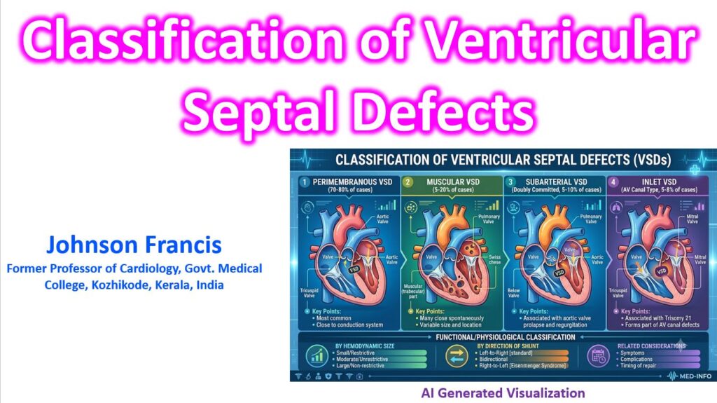

Ventricular septal defects (VSDs) are primarily classified based on their anatomical location within the ventricular septum—the wall separating the heart’s right and left lower chambers. Because the septum has a complex, 3D structure with membranous and muscular components, different naming conventions exist (such as the Anderson or STS/EACTS systems), but they all generally divide VSDs into four main types.
Anatomical Classification
The anatomical location dictates both the clinical risks (like damage to adjacent valves or the conduction system) and the approach to closure.
1. Perimembranous VSD (Type 2)
- Prevalence: The most common type, accounting for roughly 75% to 80% of all VSDs.
- Location: Located in the membranous portion of the septum, just below the aortic valve, and often extending into the adjacent muscular tissue.
- Clinical Note: Because of their proximity to the bundle of His (part of the heart’s electrical conduction system), surgical closure carries a slight risk of inducing a heart block. They can partially or fully close spontaneously if the tricuspid valve tissue forms a “pouch” or aneurysm over the defect.
2. Muscular VSD (Type 4)
- Prevalence: Accounts for up to 20% of VSDs (especially common in infants, though many close spontaneously before adulthood).
- Location: Bordered entirely by the myocardium (heart muscle) in the lower, trabeculated part of the septum.
- Subtypes: Can be further described by their specific location (anterior, mid-muscular, apical, or posterior). If multiple muscular defects are present, it is often referred to as a “Swiss cheese” septum.
3. Inlet VSD (Type 3)
- Prevalence: Accounts for 5% to 8% of VSDs.
- Location: Located posterior and inferior to the membranous septum, sitting directly beneath the inlet valves (specifically the septal leaflet of the tricuspid valve).
- Clinical Note: These defects are part of the atrioventricular (AV) septal defect spectrum and are frequently seen in patients with Down syndrome. Because they involve the structural crux of the heart, they rarely close spontaneously.
4. Outlet / Supracristal VSD (Type 1)
- Prevalence: Accounts for 5% to 7% of VSDs in Western populations, but is notably more common in Asian populations (up to 30%).
- Location: Located in the outflow tract of the right ventricle, above the crista supraventricularis (a muscular ridge). They sit right below the semilunar valves (pulmonary and aortic). Also known as doubly committed subarterial VSDs.
- Clinical Note: Because they are situated immediately below the aortic valve, the lack of support can cause the aortic valve leaflet (usually the right coronary cusp) to prolapse into the defect, leading to aortic regurgitation.
Hemodynamic Classification
Beyond anatomy, VSDs are also classified by their physiological impact, determined by the size of the defect and the resulting pressure gradient between the ventricles:
| Classification | Defect Size | Hemodynamic Impact |
| Restrictive | Small | The defect resists flow; significant pressure difference remains between the left and right ventricles. Usually asymptomatic. |
| Moderately Restrictive | Medium | Some resistance to flow, but pulmonary pressure is elevated. Can lead to left heart volume overload. |
| Unrestrictive | Large | No resistance to flow; pressures in the left and right ventricles equalize. Results in severe pulmonary hypertension and risk of Eisenmenger syndrome if uncorrected. |