


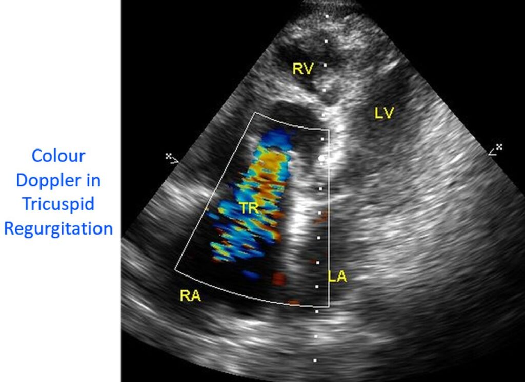

Colour Doppler in tricuspid regurgitation
Tricuspid regurgitation can be broadly divided into hypertensive and non-hypertensive, depending on the right ventricular pressure, whether it is elevated or not. Tricuspid regurgitation is best assessed in the apical four chamber view on echocardiography. This is because the direction of the jet and the Doppler beam are parallel to each other in this view. Hence the estimation of the jet velocity will be more accurate in this view. The more distally the jet extends from the tricuspid valve into the right atrium, the more severe the regurgitation. A larger jet area on colour Doppler also indicates a severe regurgitation.
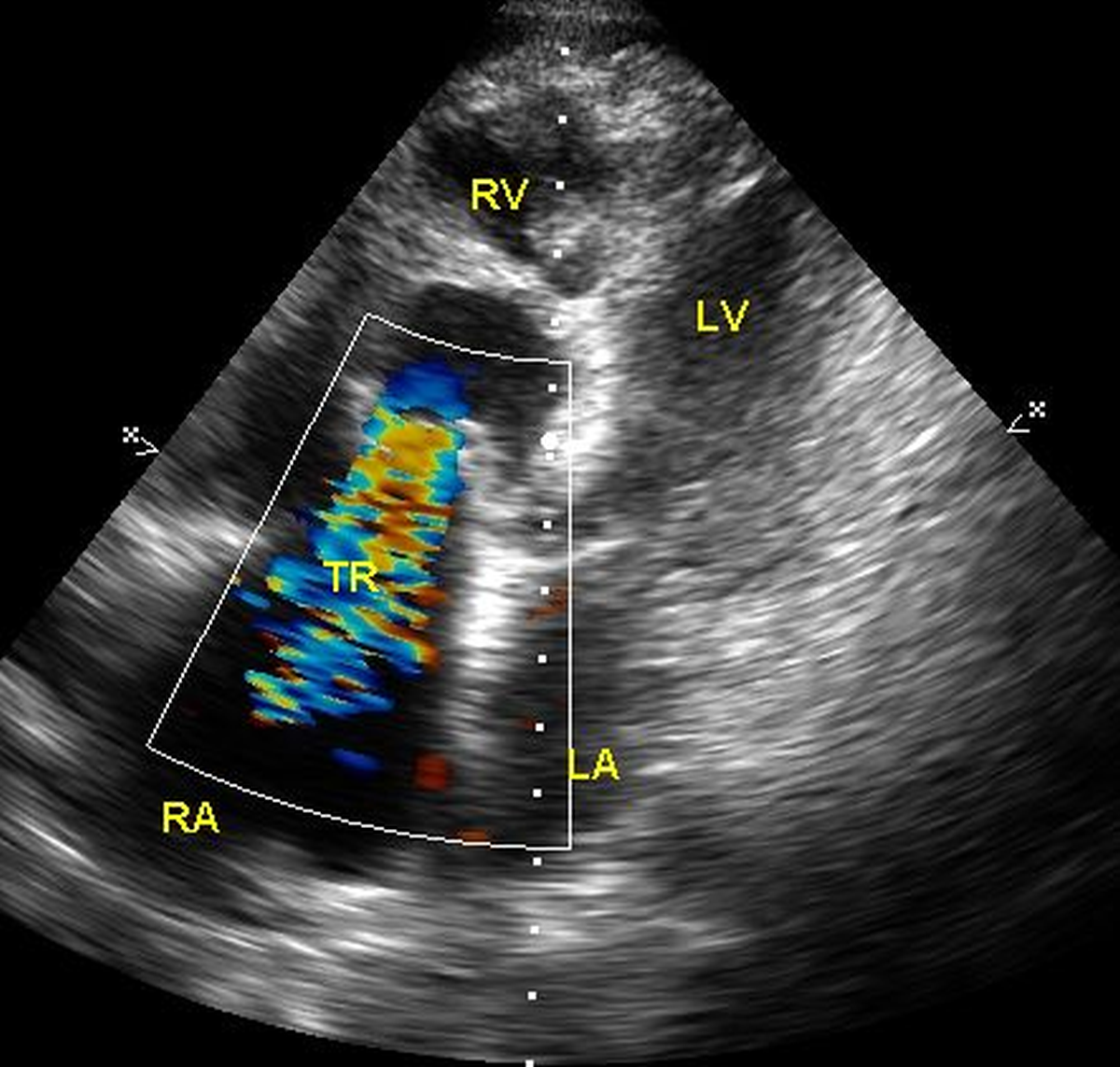

If isolated severe tricuspid regurgitation is noted without pulmonary hypertension a close look at the valve leaflets for any vegetations suggesting infective endocarditis is needed.