


The fundamental difference between Pulsed Wave (PW) and Continuous Wave (CW) Doppler lies in how the ultrasound signals are transmitted and received, which dictates their ability to pinpoint the location of blood flow versus their ability to measure high-velocity blood flow. Here is the breakdown of how each modality functions and where they are most effectively deployed in clinical echocardiography.
Pulsed Wave (PW) Doppler
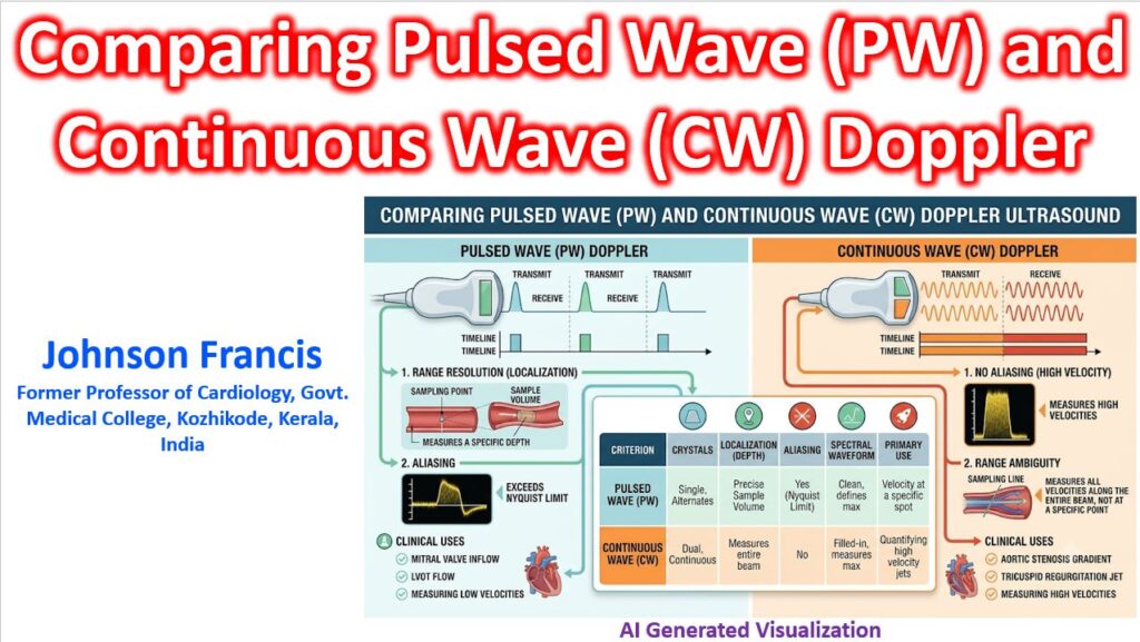
PW Doppler utilizes a single piezoelectric crystal that alternates between sending and receiving ultrasound signals. It emits a brief pulse of sound, waits for the echo to return, and then emits the next pulse.
- Range Resolution: This is the defining advantage of PW Doppler. Because the timing of the returning echo corresponds to distance, you can place a “sample volume” or “gate” at a precise depth. The machine only analyzes Doppler shifts returning from that exact location.
- The Aliasing Limitation: PW Doppler is restricted by the Nyquist limit. If the blood flow velocity is too high (typically > 1.5 to 2.0 m/s), the machine cannot sample the returning signal fast enough. The waveform “wraps around” the baseline, a phenomenon known as aliasing, making it impossible to determine the true peak velocity.
- Clinical Application: PW is used when location is critical, and velocities are normal or low. It is ideal for assessing Left Ventricular Outflow Tract (LVOT) velocities, profiling mitral valve inflow (E and A waves) for diastolic function, or mapping the precise origin of a regurgitant jet.
Continuous Wave (CW) Doppler
CW Doppler uses a transducer with two separate crystals: one continuously transmits ultrasound waves, while the other continuously receives the returning echoes.
- High-Velocity Measurement: Because transmission and reception are continuous, there is no waiting period and therefore no Nyquist limit. CW Doppler can accurately measure extremely high-velocity blood flow without aliasing.
- Range Ambiguity: The primary limitation of CW Doppler is its inability to pinpoint exactly where the velocity is occurring along the ultrasound beam. It records all Doppler shifts along the entire length of the beam.
- Clinical Application: CW is essential when precise velocity quantification takes precedence over depth localization. It is the standard for measuring peak gradients across tight, stenotic valves (like severe aortic stenosis) or calculating right ventricular systolic pressure from a high-velocity tricuspid regurgitation jet.
Key Differences at a Glance
| Feature | Pulsed Wave (PW) Doppler | Continuous Wave (CW) Doppler |
| Crystal Configuration | Single crystal (alternates send/receive) | Two crystals (one sends, one receives) |
| Depth/Location | Range Resolution: Exact depth is known | Range Ambiguity: Exact depth is unknown |
| Velocity Limits | Low limit (subject to aliasing) | No practical limit (no aliasing) |
| Primary Echo Utility | Pinpointing flow location (e.g., LVOT, pulmonary veins) | Quantifying high velocities (e.g., Aortic Stenosis, TR jets) |
| Audio/Visual Profile | “Clean” spectral envelope | “Filled-in” spectral envelope |