Continuous wave (CW) Doppler imaging in aortic stenosis
Continuous wave (CW) Doppler imaging in aortic stenosis
Aortic stenosis produces a high velocity jet of blood across the aortic valve. This high velocity jet can be imaged well only by continuous wave Doppler imaging as it will be well above the aliasing velocity for pulse Doppler. Aliasing velocity is the maximum velocity which can be imaged by a given Doppler frequency and is dependent on the pulse repetition frequency. The highest velocity that can be detected is known as the Nyquist limit.
Nyquist limit = half the pulse repetition frequency
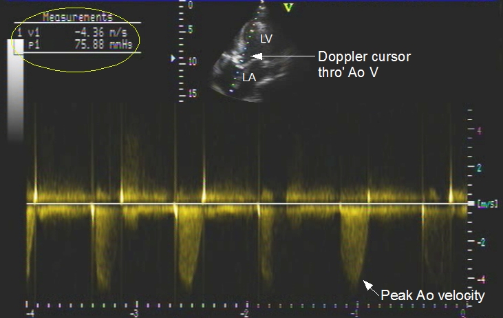
The small figure above shows the line of the CW Doppler cursor passing through the aortic valve. The lower portion shows the tongue shaped Doppler signal due to systolic flow in the aorta just beyond the aortic valve at high velocity. The scale at the bottom represents time axis and the vertical scale represents the velocity in m/sec. The scale beside the 2-D image at the top gives the depth in cm. Top left corner displays the measurements, in this case the peak aortic velocity, 4.36 m/s and gradient, 75.88 mm Hg.
The gradient is calculated from the velocity using the Bernoulli equation:
Pressure gradient = 4 V2, where V is the velocity.
The mean gradient can be estimated manually or electronically sketching out the envelope of the jet and the computer program generates the mean gradient display. The gradient in this case is quite high, representing severe aortic stenosis. A limitation of assessing the severity by gradient alone is that the gradient may be falsely low when the left ventricle fails and is unable to generate adequate pressures. This can be seen in the Doppler tracing as a less steeper slope of the tracing from the base to peak. The ventricular dp/dt can be estimated from this slope. Steeper the slope, higher the ventricular dp/dt, which is an important measure of the contractile state of the left ventricle.
Related Posts


About The Author
Johnson Francis
Former Professor of Cardiology, Calicut Govt. Medical Kozhikode, Kerala, India. Editor-in-Chief, BMH Medical Journal