


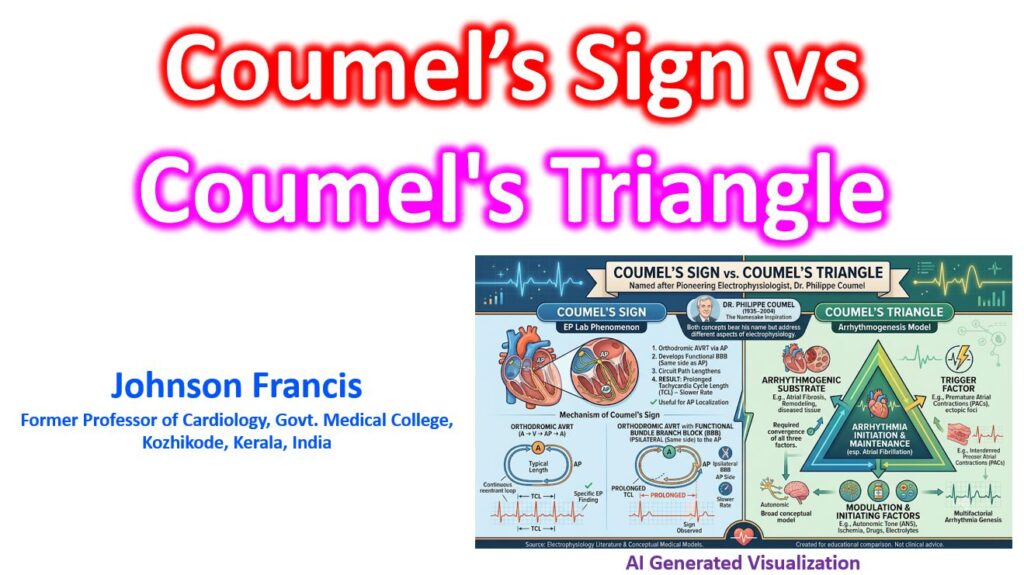

While both concepts are named after the renowned French electrophysiologist Philippe Coumel, they refer to fundamentally different aspects of cardiology. Coumel’s sign is a specific diagnostic electrocardiographic finding used to localize accessory pathways, whereas Coumel’s Triangle is a foundational pathophysiological model used to explain how arrhythmias develop.
Coumel’s Sign (Coumel’s Law)
Coumel’s sign is an electrophysiologic observation seen during orthodromic atrioventricular reentrant tachycardia (AVRT), such as in Wolff-Parkinson-White (WPW) syndrome.
It states that if a functional bundle branch block (BBB) develops on the same side (ipsilateral) as the accessory pathway during the tachycardia, the tachycardia cycle length (TCL) will increase—meaning the heart rate slows down.
- The Mechanism: In orthodromic AVRT, the electrical impulse travels down the AV node/His-Purkinje system and retrograde up the accessory pathway. If an ipsilateral BBB occurs, the impulse can no longer travel down the bundle branch on that side. Instead, it must travel down the contralateral bundle branch, cross the interventricular septum, and then travel up the accessory pathway.
- The Result: This anatomic detour increases the size of the macroreentrant circuit. It manifests as a prolongation of the ventriculoatrial (VA) interval (typically by ≥ 35 ms) and a corresponding increase in the overall TCL.
- Clinical Utility: It is highly useful in the EP lab (and on surface ECGs) to lateralize an accessory pathway. For example, if left bundle branch block (LBBB) develops during AVRT and the tachycardia slows down, it confirms the presence of a left-sided accessory pathway.
Coumel’s Triangle of Arrhythmogenesis
Coumel’s Triangle is a conceptual framework that describes the three necessary elements required for a clinical arrhythmia to initiate and sustain itself. It is most famously applied to atrial fibrillation and ventricular tachycardia.
According to Coumel, an arrhythmia is not caused by a single defect, but rather the interaction of three distinct pillars:
- The Arrhythmogenic Substrate: The underlying structural or electrophysiological abnormality in the myocardium. This could be myocardial scar tissue from a prior infarction, atrial fibrosis, hypertrophy, or a genetic channelopathy that allows for reentry or abnormal automaticity.
- The Trigger: The initiating electrical event that sparks the arrhythmia. This is typically an ectopic beat, such as a premature atrial contraction (PAC) originating from the pulmonary veins, or a premature ventricular contraction (PVC).
- The Modulating Factors: The dynamic, transient influences that alter the electrophysiological properties of the heart, making the substrate susceptible to the trigger. The most prominent modulator is the autonomic nervous system (fluctuations in sympathetic or parasympathetic/vagal tone), but it can also include acute ischemia, electrolyte imbalances, or drug toxicity.
- Clinical Utility: It forms the basis of arrhythmia management. Therapy aims to address one or more sides of the triangle—such as ablating the substrate/trigger, or using beta-blockers to alter the autonomic modulator.
Summary Comparison
| Feature | Coumel’s Sign (Law) | Coumel’s Triangle |
| Concept Type | Diagnostic electrocardiographic finding | Pathophysiological framework/model |
| Primary Application | Orthodromic AVRT (WPW syndrome) | All clinical arrhythmias (especially AFib/VT) |
| Core Principle | Ipsilateral BBB increases the VA interval and slows tachycardia cycle length. | Arrhythmias require the interaction of a substrate, a trigger, and a modulator. |
| Clinical Goal | To localize the anatomical side of an accessory pathway. | To guide therapeutic strategies by targeting the specific mechanisms of an arrhythmia. |
References
- Coumel P., Attuel P. Reciprocating tachycardia in overt and latent preexcitation. Influence of functional bundle branch block on the rate of the tachycardia. Eur J Cardiol. 1974;1:4: 423-436.
- Coumel P. The management of clinical arrhythmias. An overview on invasive versus non-invasive electrophysiology. Eur Heart J. 1987; 8:92–99.
- Coumel P, Leenhardt A. Mental activity, adrenergic modulation, and cardiac arrhythmias in patients with heart disease. Circulation. 1991; 83(4 Suppl): I58–170.
- Coumel P. Cardiac arrhythmias and the autonomic nervous system. J Cardiovasc Electrophysiol. 1993; 4:338–355.