


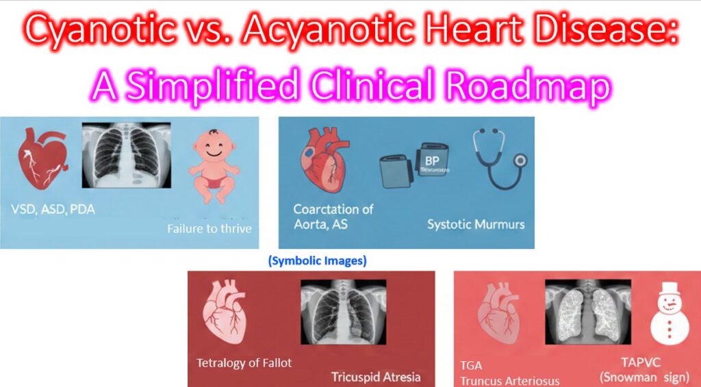

Understanding congenital heart disease is often easier when you move away from exhaustive lists and focus on the hemodynamic logic. Here is a simplified clinical roadmap to help differentiate and diagnose these conditions based on their physiological impact.
The Acyanotic Group (Left-to-Right Shunts Plus)
In the left to right shunts, oxygenated blood from the left side of the heart leaks back into the right side. The child is “pink,” but the lungs are over circulated.
| Category | Primary Examples | Key Finding |
| Volume Overload in shunts | VSD, ASD, PDA | Increased pulmonary markings on X-ray; failure to thrive. |
| Pressure Overload in obstructions | Coarctation of Aorta, AS | Discrepancy in upper vs. lower limb pulses in coarctation; systolic murmurs. |
- The Clinical Logic: The main issue here isn’t oxygen saturation—it’s congestive heart failure (CHF). If the shunt is large, the heart works too hard to pump the extra volume, leading to tachypnea and poor feeding. Coarctation and aortic stenosis can also lead to heart failure if severe enough.
The Cyanotic Group (Right-to-Left Shunts)
Here, deoxygenated blood bypasses the lungs and enters systemic circulation. The child is “blue.” To simplify these, look at the Pulmonary Blood Flow (PBF) on a chest X-ray.
A. Decreased PBF (The “Dark Lungs” Group)
The blood can’t get to the lungs because of an obstruction.
- Tetralogy of Fallot (ToF): The most common. Characterized by the “boot-shaped” heart.
- Tricuspid Atresia: No valve, so blood must cross an ASD to survive.
B. Increased PBF (The “Wet Lungs” Group)
Blood is mixing, but there is no obstruction to the lungs.
- Transposition of the Great Arteries (TGA): The “egg-on-a-string” heart. This is a neonatal emergency.
- Truncus Arteriosus: One single vessel supplying both the body and lungs.
- TAPVC: Total Anomalous Pulmonary Venous Connection (“Snowman” sign).
The Quick Diagnostic Algorithm
When you see a suspected CHD case, ask these three questions in order:
- Is the patient cyanotic? * No: Think VSD, ASD, or Coarctation.
- Yes: Proceed to step 2.
- What does the X-ray show?
- Clear/Dark Lungs: Think ToF (obstruction to pulmonary flow).
- Congested/White Lungs: Think TGA or Truncus (mixing with high flow).
- Is the EKG showing Right or Left Axis Deviation?
- Left Axis + Cyanosis: Highly suggestive of Tricuspid Atresia.
- Right Axis + Cyanosis: Common in ToF or TGA.