


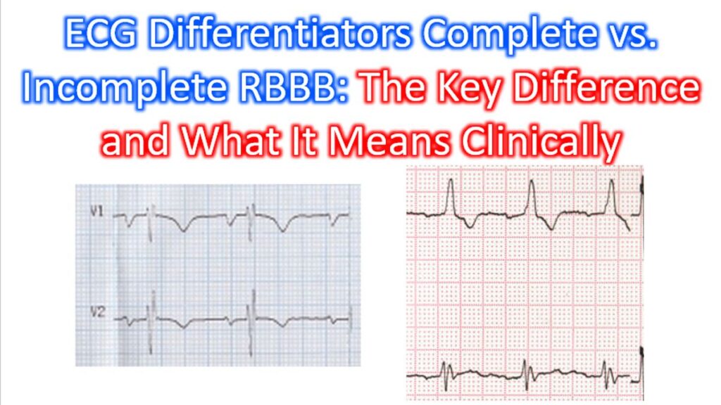

Understanding the distinction between Complete and Incomplete Right Bundle Branch Block (RBBB) is essential for accurate ECG analysis. While they share a common “look,” the difference lies entirely in the speed of conduction through the right ventricle.
1. The Key Diagnostic Differentiator: QRS Duration
The primary way to distinguish between the two is the measurement of the QRS duration. Both patterns involve a delay in right ventricular depolarization, but the extent of that delay defines the category.
| Feature | Incomplete RBBB | Complete RBBB |
| QRS Duration | Between 100 ms and 120 ms | ≥ 120 ms (0.12s) |
| V1 Morphology | rsr’, rSR’, or rsR’ | rsr’, rSR’, or rsR’ (M-shape) |
| Lead I / V6 | Shallow, slurred S wave | Wide, deep, slurred S wave |
Morphological Similarities
In both cases, you will see the classic “Rabbit Ears” in the right precordial leads (V1-V2). This happens because the left ventricle depolarizes first (the ‘r’), followed by a delayed signal traveling to the right ventricle (the ‘R”).
2. Electrophysiology: What is Happening?
The Right Bundle Branch (RBB) is a long, thin, and fragile part of the conduction system.
- Complete RBBB: The electrical impulse is entirely blocked in the RBB. The right ventricle must wait for the impulse to travel through the left bundle and then “seep” through the myocardium (muscle-to-muscle conduction). This is a slow process, hence the QRS width ≥ 120 ms.
- Incomplete RBBB: The RBB is not completely blocked but conducts more slowly than usual, or there is mild right ventricular hypertrophy (RVH) causing a slight delay. The signal eventually gets through the conduction system, but it takes just a bit longer than the standard 100 ms.
3. Clinical Significance
The implications of these findings vary significantly based on the patient’s presentation.
Incomplete RBBB
- Normal Variant: Often found in healthy, asymptomatic individuals (especially athletes) with no underlying heart disease.
- Right Ventricular Strain: Can be a sign of early or mild RVH, often seen in conditions like Atrial Septal Defect (ASD).
- Progression: It rarely progresses to a complete heart block on its own.
Complete RBBB
- Pathological Associations: More frequently associated with structural heart disease, such as coronary artery disease, hypertensive heart disease, or pulmonary embolism (acute onset).
- Diagnostic Interference: Unlike Left Bundle Branch Block (LBBB), a Complete RBBB does not usually hide an acute myocardial infarction (STEMI) on an ECG, as the initial phase of the QRS (septal depolarization) remains intact.
- Surgical Context: New-onset RBBB is a common occurrence following certain cardiac surgeries.
Summary Checklist
When you see an rSR’ pattern in V1:
- Check the width: Is it <120 ms? (Incomplete) or ≥120 ms? (Complete).
- Check the S-wave: Is there a slurred S-wave in Lead I or V6? (Confirms RBBB).
- Check the Axis: Is there a concomitant Axis Deviation? (Consider Bifascicular Block).