


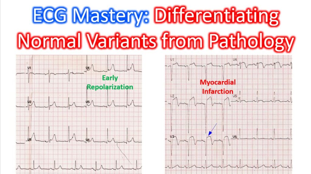

Differentiating between benign ECG variants and true pathology is one of the most important skills in clinical cardiology. In a high-volume practice, misinterpreting a normal variant as pathology leads to unnecessary anxiety and testing, while missing a subtle pathological sign can be catastrophic.
Here is a breakdown of the most common “look-alikes” encountered in clinical practice.
1. ST-Segment Elevation: BER vs. STEMI
Benign Early Repolarization (BER) is perhaps the most frequent mimic of an acute MI.
| Feature | Benign Early Repolarization (BER) | Acute STEMI |
| ST Morphology | Concave (“smiley face”) | Convex or straight (“tombstoning”) |
| J-point | Notched or slurred (the “fishhook”) | Elevated without distinct notching |
| Reciprocal Changes | Absent (except in aVR) | Present (highly specific) |
| T-waves | Large, symmetrical, concordant | Hyperacute or inverted (evolutionary) |
| Stability | Remains stable over time | Dynamic changes within minutes |
Clinical Pearl: If you see ST elevation in V2–V5 with a notched J-point and no reciprocal depression in the inferior leads, think BER. If the ST segment is convex and there is even 0.5mm of depression in III or aVF, treat as STEMI until proven otherwise.
2. The “Athletic Heart” vs. Cardiomyopathy
Athletes often exhibit ECG changes that would be highly concerning in a sedentary patient. The International Criteria (2017) help distinguish these.
Normal Variants in Athletes
- Sinus Bradycardia: Often <40 bpm; normal if asymptomatic.
- First-Degree AV Block: Common due to high vagal tone.
- Incomplete RBBB: Very common; does not require workup.
- Early Repolarization: High-voltage QRS with ST elevation.
Red Flags (Pathology)
- T-Wave Inversion: Deep T wave inversion in V4–V6, I, or aVL is a major red flag for HCM.
- ST-Segment Depression: Never a normal variant in an athlete.
- Pathological Q-waves: Q/R ratio ≥0.25 or ≥40 ms in duration in two or more contiguous leads (except III and aVR).
3. Right Ventricular Patterns: Brugada vs. Others
The “Saddleback” or “Coved” ST elevation in V1–V2 can be terrifying but isn’t always Brugada Syndrome.
- Brugada Type 1: Coved ST elevation >2 mm followed by a negative T-wave in V1–V2. This is diagnostic.
- Brugada Type 2 (Saddleback): ST elevation >2 mm but with a positive or biphasic T-wave. This can be a normal variant or related to pectus excavatum. But if this pattern is converted to type 1 by drug challenge with medications like flecainide, it is indicative of Brugada syndrome. Please note that drug challenge should be done only with great precaution as it can rarely precipitate serious ventricular arrhythmias.
4. T-Wave Inversions: When to Worry or not!
Not all flipped T-waves mean ischemia.
- Persistent Juvenile Pattern: T-wave inversion in V1–V3 in a young, asymptomatic adult (more common in females).
- Wellens Syndrome: Deeply inverted or biphasic T-waves in V2–V3 while the patient is chest-pain free. This represents a critical LAD stenosis—a true “pre-infarction” state.
- Cerebral T-waves: Massive, wide, deeply inverted T-waves seen in intracranial hemorrhage or massive stroke.