

ECG Quiz 3

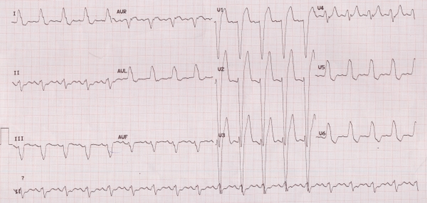
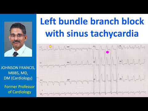
Wide QRS is due to left bundle branch block as evidenced by the negative QRS complex in lead V1 and slurred R waves in I, aVl and V5, V6. Secondary ST segment and T wave changes are also seen, opposite in direction to the dominant QRS – ST elevation with upright T waves in right precordial leads and ST segment depression with inverted T waves in left precordial leads. If the ST segment and T waves are concordant, it will indicate a primary T wave abnormality as in myocardial ischemia. Left ward axis is considered by some authors as evidence of additional left anterior fascicular block.