ECG Quiz 42
ECG Quiz 42
Discussion
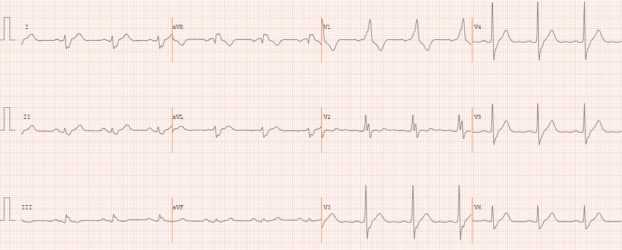
QRS axis has shifted to the right indicating right posterior hemiblock. rS complexes are seen in I and aVL. Wide QRS with notched R in V1, V2 is suggestive of right bundle branch block. The slurred S waves in I and aVL are also due to RBBB. PR interval is 200 ms. RBBB + LPHB constitutes one form of bifascicular block. Left posterior fascicular block is rarer because it is a thicker fascicle with dual blood supply. Hence presence of LPHB indicates more advanced disease.
Related Posts


About The Author
Johnson Francis
Former Professor of Cardiology, Calicut Govt. Medical Kozhikode, Kerala, India. Editor-in-Chief, BMH Medical Journal
One Comment
Sinus rhythm,60/min
Complete RBBB
Low voltage ECG
S1Q3T3
Right axis