

ECG Quiz 42

Discussion
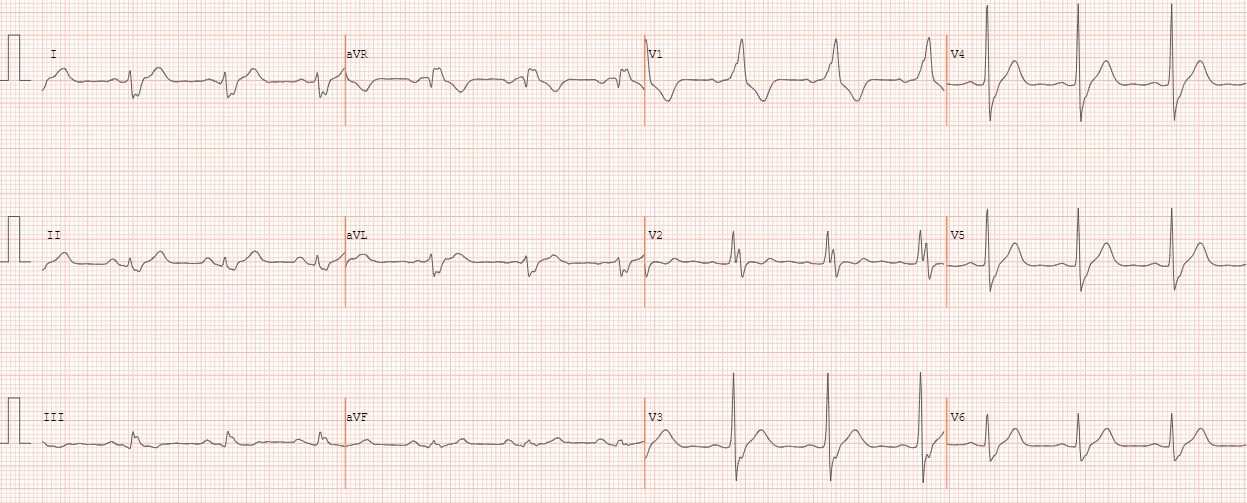
QRS axis has shifted to the right indicating right posterior hemiblock. rS complexes are seen in I and aVL. Wide QRS with notched R in V1, V2 is suggestive of right bundle branch block. The slurred S waves in I and aVL are also due to RBBB. PR interval is 200 ms. RBBB + LPHB constitutes one form of bifascicular block. Left posterior fascicular block is rarer because it is a thicker fascicle with dual blood supply. Hence presence of LPHB indicates more advanced disease.