Echocardiogram in PLAX view with video
Echocardiogram in PLAX view with video
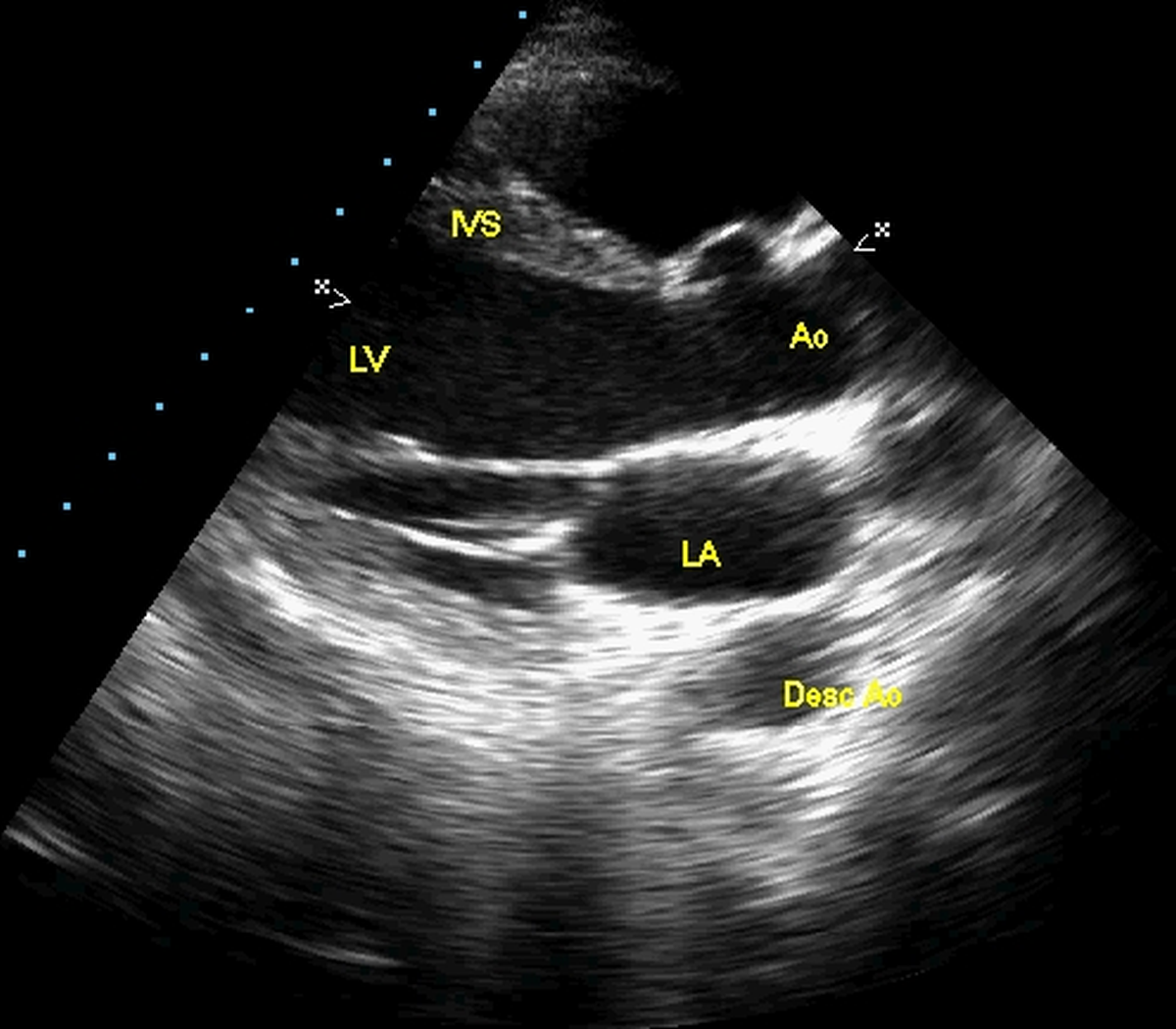
Echocardiogram still frame in parasternal long axis view demonstrating IVS: interventricular septum (with right ventricle above – not marked); Ao: aorta (root of aorta); LA: left atrium; Desc Ao: descending aorta in cross section; LV: left ventricle
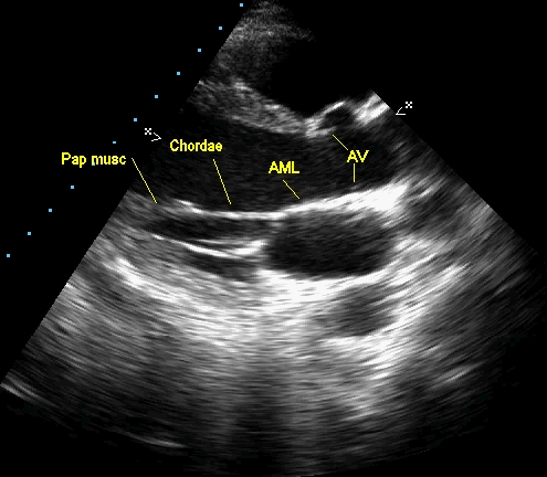
Same view repeated for adding more annotations: AML: anterior mitral leaflet (closed position); AV: aortic valve leaflets (open position); Chordae: chordae tendinae attached to AML; Pap musc: papillary muscle of the left ventricle, attached to the chordae of AML; Posterior papillary muscle and chordae are seen just below that.
Related Posts


About The Author
Johnson Francis
Former Professor of Cardiology, Calicut Govt. Medical Kozhikode, Kerala, India. Editor-in-Chief, BMH Medical Journal