Echocardiogram in mitral stenosis and regurgitation
Echocardiogram in mitral stenosis and regurgitation
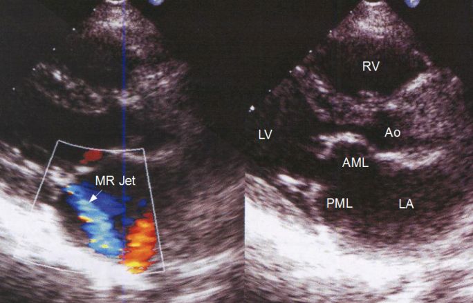
Echocardiographic image in parasternal long axis view illustrating a combination of mitral stenosis and regurgitation. Right half shows the two dimensional image while the left half shows superimposed colour Doppler image. The bluish mosaic coloured jet within the quadrangular Doppler sample volume is suggestive of mild mitral regurgitation. The left atrium is dilated, when compared to the aorta, both of which are usually of equal size in this view in normals. Left ventricular size is normal. The anterior mitral leaflet is seen doming into the left ventricle in diastole. The posterior mitral leaflet is pulled along with it due to commissural fusion, causing the paradoxical anterior motion in diastole. Normally the anterior leaflet moves anteriorly and posterior leaflet moves posteriorly while opening in ventricular diastole, allowing free blood flow from the left atrium to the left ventricle. Both doming of the anterior mitral leaflet and paradoxical anterior motion of the posterior mitral leaflet are characteristic echocardiographic features of rheumatic mitral stenosis.
Related Posts

About The Author
Johnson Francis
Former Professor of Cardiology, Calicut Govt. Medical Kozhikode, Kerala, India. Editor-in-Chief, BMH Medical Journal