Echocardiogram in Severe Pulmonary Hypertension
Echocardiogram in severe pulmonary hypertension
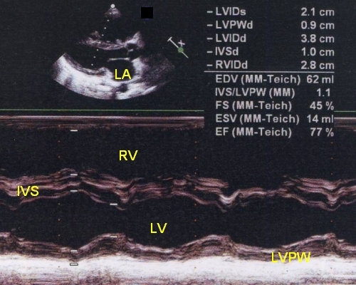
M-mode echocardiogram shows dilated right ventricle, mainly the outflow region is seen in this view. IVS: interventricular septum; LV: left ventricle; LVPW: left ventricular posterior wall; LA: left atrium; LVIDs: left ventricular internal diameter, systolic; LVPWd: left ventricular posterior wall, diastolic; LVIDd: left ventricular internal diameter, diastolic; IVSd: interventricular septum, diastolic; EDV: end diastolic volume; FS: fractional shortening; ESV: end systolic volume; EF: ejection fraction; IVS/LVPW: septal to posterior wall ratio. When the right ventricle is dilated due to volume overload as in atrial septal defect, the septal motion becomes paradoxical, i.e, moves towards the right ventricular free wall in systole. Here the septal motion is towards the left ventricle in systole and suggests that the right ventricular enlargement is due to pressure overload rather than volume overload. Estimation of ejection fraction by M-mode becomes erroneous if paradoxical septal motion is present.
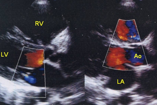
Dilated right ventricle seen on parasternal long axis view on two dimensional echocardiography with colour Doppler imaging. LA: left atrium; Ao: aorta. There is a trivial mitral regurgitation jet seen in the left atrium as a blue jet in the left frame, with the mitral valve in closed position, which is of no practical significance.
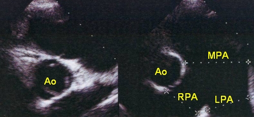
Dilated main pulmonary artery, left pulmonary artery and right pulmonary artery on short axis view of 2-D echocardiogram. Usually the aorta and main pulmonary artery are of comparable size. Here the main pulmonary artery is grossly dilated and about twice the size of aorta, due to severe pulmonary hypertension.
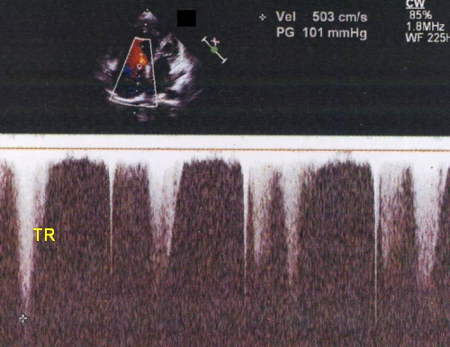
Tricuspid regurgitation gradient in severe pulmonary hypertension of 101 mm Hg. The TR jet is incompletely visualised. Estimated right ventricular systolic pressure is obtained by adding the right atrial pressure to this value, taken as 10 mm Hg nominally. As the velocity is very high interrogation is done using continuous wave Doppler.
Related Posts


About The Author
Johnson Francis
Former Professor of Cardiology, Calicut Govt. Medical Kozhikode, Kerala, India. Editor-in-Chief, BMH Medical Journal