


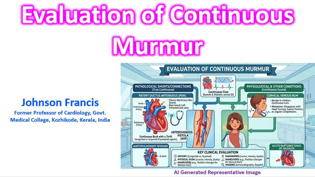

The evaluation of a continuous murmur requires a systematic approach to differentiate between benign physiological flows and complex pathological shunts. A continuous murmur is defined by its timing: it begins in systole and continues through the second heart sound (S2) into part or all of diastole without interruption.
1. Pathophysiological Mechanism
Continuous murmurs are generated when there is a persistent pressure gradient between two cardiovascular structures or vessels throughout the cardiac cycle. This results in uninterrupted blood flow from a high-pressure or high-resistance system to a low-pressure or low-resistance system.
It is vital to distinguish a “true continuous” murmur from a “to-and-fro” murmur. In a to-and-fro murmur (e.g., combined aortic stenosis and regurgitation), blood flows in opposite directions during systole and diastole. In a true continuous murmur, blood maintains the same direction of flow across both phases.
2. Differential Diagnosis by Etiology
The causes of continuous murmurs are broadly categorized based on the site of the shunt or the nature of the vessel involved.
High-to-Low Pressure Shunts
- Patent Ductus Arteriosus (PDA): The most classic cause. It produces a “machinery-like” murmur loudest at the left infraclavicular area or second left intercostal space. The systolic component peaks at S2 and continues into diastole. The murmur is also known as Gibson’s murmur and train in tunnel murmur.
- Ruptured Sinus of Valsalva Aneurysm: Often presents with sudden onset chest pain or dyspnea. The murmur is typically loudest at the lower left sternal border and may radiate to the xiphoid process. Diastolic accentuation is a classic feature of RSOV into RV as the track gets compressed in systole, reducing the intensity of systolic component.
- Coronary Artery Fistula: A rare anomaly where a coronary artery communicates with a heart chamber or the pulmonary artery. The location of the murmur varies depending on the drainage site (e.g., right atrium vs. pulmonary trunk). A fistula communicating to a cardiac chamber is called coronary cameral fistula.
Physiological & Extracardiac Flows
- Cervical Venous Hum: A common benign finding in children and some young adults. It is best heard in the right supraclavicular fossa. It is highly position-dependent; it typically disappears when the patient lies supine or with digital compression of the internal jugular vein.
- Mammary Soufflé: Occurs during late pregnancy or the postpartum period due to increased blood flow to the breasts. It is loudest over the breasts in systole and diastole and can be abolished by firm pressure with the stethoscope.
Arterial & Venous Obstructions
- Severe Arterial Stenosis: High-grade stenosis (typically >80%) in vessels like the renal, carotid, or femoral arteries can generate a continuous bruit if the pressure gradient remains high throughout diastole.
- Cruveilhier-Baumgarten Syndrome: A continuous venous hum heard in the epigastric region due to dilated paraumbilical veins in the setting of portal hypertension.
3. Diagnostic Approach and Maneuvers
Physical examination findings and dynamic maneuvers are the first steps in narrowing the differential.
Physical Examination Keys
| Feature | PDA | Ruptured Sinus of Valsalva | Venous Hum |
| Maximal Intensity | LUSB / Left Infraclavicular | Lower LSB / Xiphoid | Right Supraclavicular |
| Pulse Quality | Bounding (Wide pulse pressure) | Collapsing | Normal |
| Maneuver Effect | Little change with position | Intensified by Handgrip | Disappears in Supine position |
Investigative Pathway
- Electrocardiogram (ECG): May show evidence of chamber enlargement (e.g., left ventricular hypertrophy in PDA).
- Chest X-Ray: Can identify pulmonary plethora or specific contours (e.g., prominent pulmonary artery).
- Transthoracic Echocardiography (TTE) with Doppler: The gold standard for initial evaluation. It confirms the presence of the shunt, determines its direction, and assesses hemodynamic impact.
- Advanced Imaging (CT/MRI/TEE): Indicated for complex anatomies like coronary-pulmonary fistulas or when TTE windows are inadequate.