


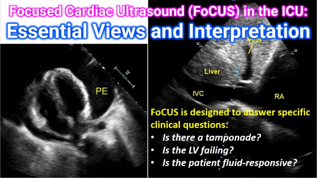

Focused Cardiac Ultrasound (FoCUS) is a simplified, goal-directed point-of-care ultrasound (POCUS) examination performed by ICU clinicians. Unlike a comprehensive echocardiogram, FoCUS is designed to answer specific clinical questions: Is there a tamponade? Is the LV failing? Is the patient fluid-responsive?
1. The 5 Essential Views
A standard FoCUS exam consists of five primary views. Each provides a unique “slice” of the heart to evaluate function and anatomy.
| View | Probe Position | Key Structures Visible | Primary Purpose |
| Parasternal Long Axis (PLAX) | 3rd–4th intercostal space (ICS), left of sternum. Marker to R shoulder. | LV, LA, RV (outflow), Aortic & Mitral valves. | Global LV function, aortic root size, pericardial effusion. |
| Parasternal Short Axis (PSAX) | Rotate probe 90° clockwise from PLAX. Marker to L shoulder. | LV (cross-section), RV. | Regional wall motion, LV “doughnut” shape, RV pressure (D-sign). |
| Apical 4-Chamber (A4C) | Point of Maximal Impulse (PMI) / 5th ICS. Marker to L axilla. | LV, RV, LA, RA, Septum. | Comparing RV vs. LV size, assessing contractility and valvular flow. |
| Subcostal (Subxiphoid) | Below xiphoid process, angled toward L shoulder. | All 4 chambers (through the liver). | Best view for pericardial effusion and tamponade. |
| Inferior Vena Cava (IVC) | Subcostal, rotate 90° clockwise. Marker toward head. | IVC entering the Right Atrium. | Assessing volume status and fluid responsiveness. |
2. Interpretation: Key Clinical Findings
In the ICU, interpretation focuses on “binary” or qualitative assessments (e.g., “Yes” vs. “No”).
Left Ventricular (LV) Function
- Hyperdynamic: LV walls almost touch in systole (“kissing papillary muscles”). Often seen in hypovolemia or distributive shock (sepsis).
- Severely Depressed: Minimal wall thickening or inward movement. Suggests cardiogenic shock or acute-on-chronic heart failure.
Right Ventricular (RV) Strain
- RV Dilation: Normally, the RV should be < 2/3 the size of the LV. If the RV is larger than the LV, suspect Pulmonary Embolism (PE) or acute RV failure.
- The “D-Sign”: In PSAX, if the interventricular septum is flattened (making the LV look like a “D” instead of a “O”), it indicates RV pressure/volume overload.
Pericardial Effusion & Tamponade
- Effusion: Anechoic (black) space around the heart.
- Tamponade Signs: Right atrial collapse in diastole or right ventricular collapse in early diastole. This is a medical emergency requiring immediate intervention.
Volume Status (IVC Assessment)
- Plethoric IVC: (Diameter > 2.1 cm with < 50% collapse) suggests high CVP (fluid overload, tamponade, or RV failure).
- Collapsible IVC: (Diameter < 2.1 cm with > 50% collapse) suggests the patient may be fluid-responsive in the setting of shock.
3. Integration into ICU Protocols
FoCUS is rarely used in isolation. It is typically part of broader diagnostic algorithms:
- RUSH Protocol: (Rapid Ultrasound in Shock) – Evaluates “The Pump” (Heart), “The Tank” (IVC/Fluids), and “The Pipes” (Aorta/DVT).
- FATE Protocol: (Focus Assessed Transthoracic Echocardiography) – A systematic approach to rule out the most common causes of circulatory failure.
Note: FoCUS is a screening tool. If you find significant valvular disease or complex pathology, a formal Comprehensive Echocardiogram by a cardiologist is indicated.