

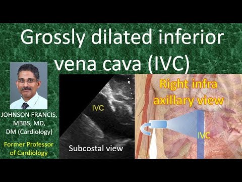
Grossly dilated inferior vena cava (IVC)
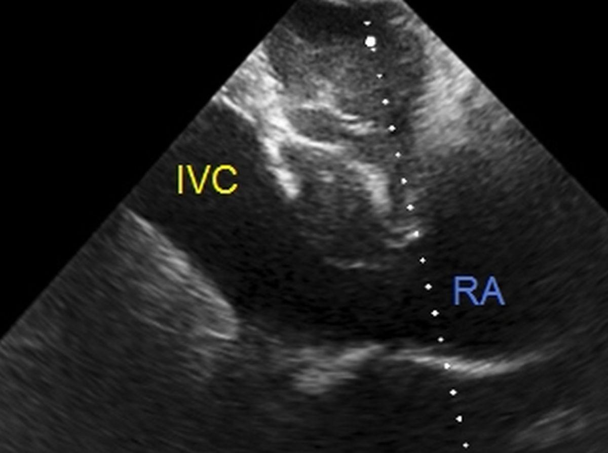

IVC measurement is often used to guide fluid resuscitation in the acute care setting instead of the conventional measurement of central venous pressure by a catheter. This is convenient being non-invasive and easily available in modern day emergency departments and critical care/intensive care units equipped with a point of care ultrasound machine. In a person presenting with hypotension, if IVC is collapsed, a fluid bolus can be given fast, often with rapid recovery.
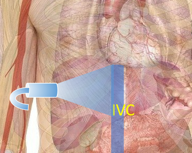
Post surgical patients will need different approach to imaging IVC as the epigastric region may be covered with surgical dressing. In these cases, imaging through the liver from right infra axillary region is very useful, though it is often a bit tricky and needs experience to get a good view of the IVC. Luboch M and colleagues evaluated the inferior vena cava/aorta index measured with the transducer placed in the anterior median line and right anterior axillary line [1]. They concluded that measurements from both sites can be used interchangeably in determining the body fluid status.

Reference
- Luboch M, Łoś M, Szmygel Ł, Kosiak W. Sonographic assessment of the inferior vena cava/aorta index measured with the transducer placed in the anterior median line and right anterior axillary line – a comparison. J Ultrason. 2014 Sep;14(58):280-6.