


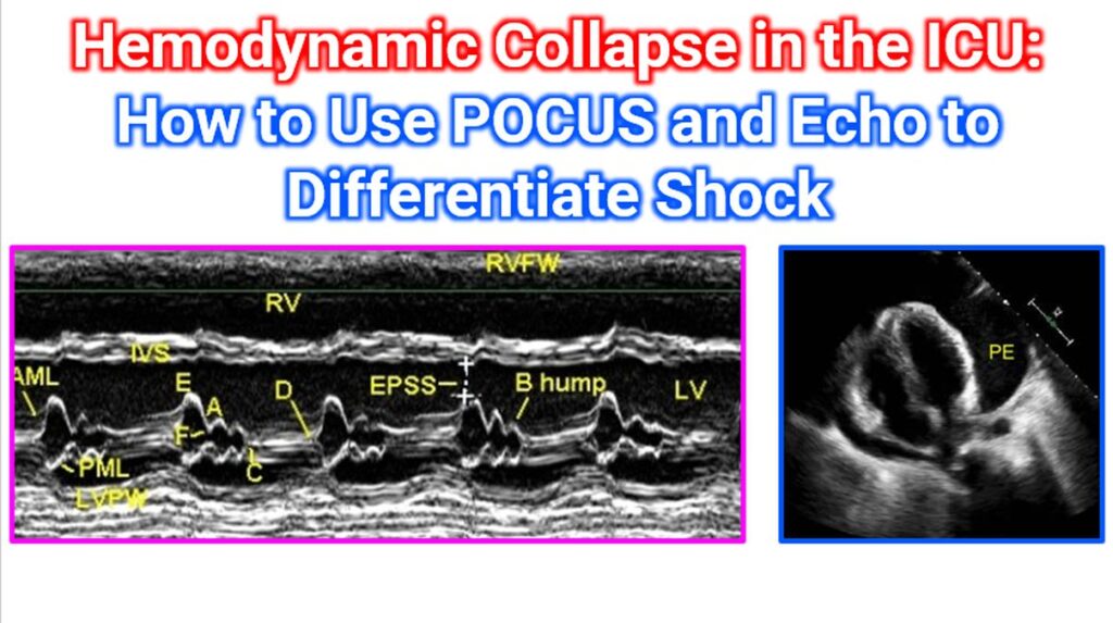

In the ICU, hemodynamic collapse requires rapid differentiation to avoid giving the wrong treatment (e.g., giving fluids to a patient in cardiogenic shock). Point-of-Care Ultrasound (POCUS) and echocardiography provide a non-invasive “window” into the patient’s physiology, allowing you to categorize shock into four main types: Hypovolemic, Cardiogenic, Obstructive, and Distributive.
The most common framework for this is the RUSH Protocol (Rapid Ultrasound for Shock and Hypotension), which systematically evaluates the Pump, the Tank, and the Pipes.
1. The Pump: Cardiac Assessment
Using the Parasternal Long Axis (PLAX), Parasternal Short Axis (PSAX), and Apical 4-Chamber (A4C) views, you can assess the heart’s function and structure.
| Shock Type | POCUS Findings (Heart) | Specific Signs |
| Hypovolemic | Hyperdynamic (vigorous contraction) | “Kissing papillary muscles” (LV walls touch in systole) |
| Cardiogenic | Hypodynamic (poor LV/RV contractility) | Dilated LV, poor mitral valve excursion (EPSS > 7mm) |
| Obstructive | RV Strain / Tamponade signs | D-sign (septal flattening), RV > LV size, Pericardial effusion |
| Distributive | Often hyperdynamic (early) | Can show late-stage sepsis-induced cardiomyopathy |
M-Mode echocardiogram showing EPSS or E point septal separation of the mitral valve echo. E point is the maximum opening motion of the anterior mitral leaflet. Normally it will reach almost up to the septum, with only minimal separation. Increased EPSS means mitral valve opening excursions are low due to low cardiac output state. This M-Mode also shows poor left ventricular contraction as evidenced by little movement of IVS towards LVPW. B hump in the AML motion indicates high left ventricular end diastolic pressure in heart failure. Pericardial effusion marked as PE appears as echo free space around the heart.
2. The Tank: Volume and Effusions
The “Tank” assessment looks at the Inferior Vena Cava (IVC) for volume status and “leaks” (free fluid).
- The IVC:
- Flat IVC (<1.5 cm) + High Collapsibility: Suggests Hypovolemic or Distributive shock.
- Distended IVC (>2.1 cm) + No Collapse: Suggests Cardiogenic or Obstructive (tamponade/pulmonary embolism) shock.
- The “Leaks” (E-FAST – Extended Focused Assessment with Sonography in Trauma):
- Check Morrison’s Pouch, the Splenorenal space, and the Pelvis for free fluid (hemorrhage).
- Lungs: Check for B-lines (pulmonary edema, typical of Cardiogenic shock) vs. Lung Sliding (absent in pneumothorax—a cause of Obstructive shock).
3. The Pipes: Great Vessels
This step identifies catastrophic vascular failures.
- Aorta: Scan for Abdominal Aortic Aneurysm (AAA) or intimal flaps (Aortic Dissection).
- Veins: Perform a two-point compression scan (femoral and popliteal veins) to look for DVT, which increases the probability of Pulmonary Embolism (Obstructive shock). In the image, obstructed vein having thrombus, with no flow on Colour Doppler is seen.
Summary of Findings by Shock Type
| Feature | Hypovolemic | Cardiogenic | Obstructive (Pulmonary Embolism/ Tamponade) | Distributive (Sepsis) |
| LV Function | Hyperdynamic | Hypodynamic | Normal/Hyperdynamic | Hyperdynamic (early) |
| IVC Size | Small/Flat | Large/Fixed | Large/Fixed | Small/Flat |
| Lungs | Clear (A-lines) | B-lines (Edema) | Clear or Pneumothorax | Clear or Focal B-lines |
| Other | Free fluid in belly | Dilated LV | RV Strain / Effusion | Source of infection |
Diagnostic “Red Flags” to Watch For:
- McConnell’s Sign: Akinesia of the RV mid-free wall with sparing of the apex (in acute pulmonary embolism).
- RA/RV Diastolic Collapse: The “shuddering” of the right chambers during diastole, indicating cardiac tamponade.
- EPSS (E-Point Septal Separation): Measure the distance between the mitral valve and the septum in PLAX. If >7mm, the LV function is likely severely reduced.