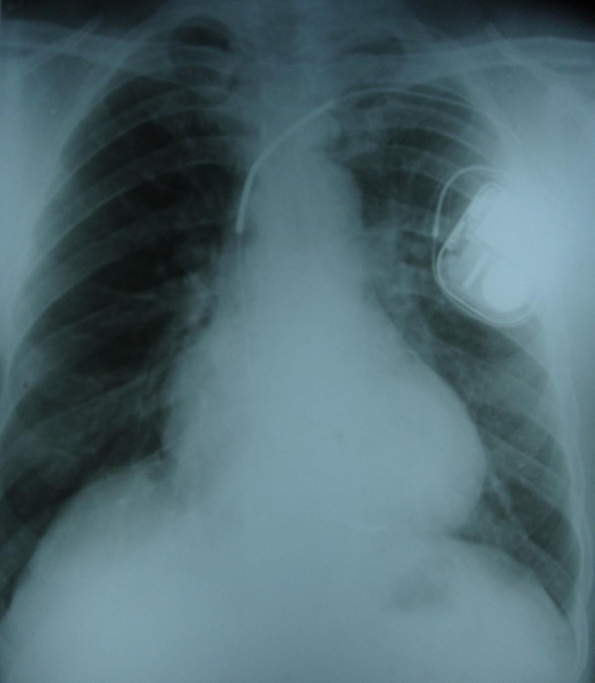
Implantable defibrillator high voltage coils on X-ray chest PA view
Implantable defibrillator high voltage coils on X-ray chest PA view
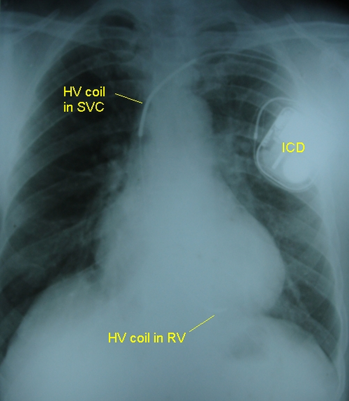
(See annotated image below)
Implantable defibrillator (ICD) high voltage coils on X-ray chest PA view. The ICD generator is seen in the left infraclavicular region . The lead is coursing through the left subclavian vein into the left brachiocephalic vein and hence into the superior vena cava. In this region the high voltage coil is seen (HV coil in SVC). Another high voltage defibrillation coil is faintly seen in the right ventricle (HV coil in RV). The ICD can is programmed as one electrode and the two high voltage coils as the other electrode. The addition of a coil in the superior vena cava reduces the defibrillation threshold compared to only a right ventricular coil. The position of the generator in the left infraclavicular region also reduces the threshold. If this position is not available, and the generator has to be implanted in the right infraclavicular region, defibrillation threshold will be high.
Related Posts


About The Author
Johnson Francis
Former Professor of Cardiology, Calicut Govt. Medical Kozhikode, Kerala, India. Editor-in-Chief, BMH Medical Journal