


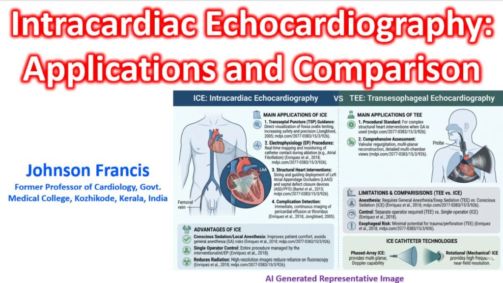

Intracardiac Echocardiography (ICE) is a highly specialized, invasive ultrasound modality that provides real-time, high-resolution imaging from inside the heart chambers. While transesophageal echocardiography (TEE) has long been the standard for guiding structural and electrophysiology (EP) procedures, ICE has increasingly become the preferred choice in modern cath labs and EP suites due to its direct visualization capabilities and patient comfort.
Key Clinical Applications
ICE is primarily used to guide complex interventional and electrophysiological procedures where precise anatomical visualization is critical.
1. Electrophysiology (EP) Procedures
- Transseptal Puncture: Provides direct visualization of the fossa ovalis, tenting of the septum by the needle, and entry into the left atrium, significantly reducing the risk of aortic or free-wall perforation.
- Atrial Fibrillation (AF) Ablation: Used to monitor catheter contact, visualize pulmonary vein anatomy, and immediately detect complications like thrombus formation or pericardial effusion.
- Ventricular Tachycardia (VT) Ablation: Helps map structural substrates and intramural scars.
2. Structural Heart Interventions
- Left Atrial Appendage Occlusion (LAAO): Used to size the appendage, guide device deployment, and assess for residual leaks.
- Septal Defect Closure: Guides the deployment of occluders for Secundum Atrial Septal Defects (ASD) and Patent Foramen Ovale (PFO).
- Transcatheter Valve Therapies: Increasingly used as an alternative to TEE during TAVR or transcatheter edge-to-edge repair (TEER) to monitor positioning and immediate hemodynamics.
ICE vs. TEE: A Quick Comparison
| Feature | Intracardiac Echo (ICE) | Transesophageal Echo (TEE) |
| Sedation | Local anesthesia / Conscious sedation | Deep sedation or General Anesthesia (GA) |
| Airway Management | Not required | Requires intubation or carries airway risks |
| Operator Control | Controlled entirely by the interventionalist/EP | Requires a separate echocardiographer |
| Imaging Artifacts | Minimal shadowing from posterior structures | Can have esophageal/airway shadowing |
| Complications | Vascular access risks, transient arrhythmias | Esophageal trauma, aspiration |
| Cost | Higher (single-use disposable catheters) | Lower (reusable probes) |
Technical Modalities
Modern ICE technology generally falls into two categories based on transducer design:
- Rotational (Mechanical) ICE: Utilizes a high-frequency (typically 9 – 12 MHz) rotating transducer that provides a 360° radial, cross-sectional view perpendicular to the catheter tip. Excellent for near-field resolution but lacks Doppler capabilities.
- Phased-Array ICE: Utilizes a lower frequency (4 – 10 MHz) linear longitudinal array providing a 90° sector beam. It supports full Color, Pulsed-Wave (PW), and Continuous-Wave (CW) Doppler, making it indispensable for evaluating flow velocities and valvular regurgitation.
- 3D/4D ICE: The latest advancement, offering real-time volumetric rendering of complex structures (like the LAA or mitral valve) without needing to manipulate the catheter manually to reconstruct the geometry.
Advantages and Limitations
Clinical Benefits
- Zero Fluoroscopy Potential: By providing continuous soft-tissue visualization, ICE significantly reduces radiation exposure for both the patient and the medical team.
- Immediate Complication Detection: Allows for instantaneous detection of pericardial effusion or thrombus formation on sheaths/wires before clinical deterioration occurs.
- Cath Lab Efficiency: Eliminates the need for an anesthesia team to manage the airway, optimizing lab turnaround times.
Limitations & Risks
- Cost: The catheters are single-use and single-patient, adding significant material cost to the procedure.
- Vascular Access: Requires an additional large-bore venous sheath (typically 8F to 11F), usually via the femoral vein.
- Learning Curve: Manipulating the catheter from the right atrium to obtain standard views (Home view, Tricuspid view, Septum view, etc.) requires specialized training.