Isorhythmic AV dissociation
ECG showing isorhythmic AV dissociation
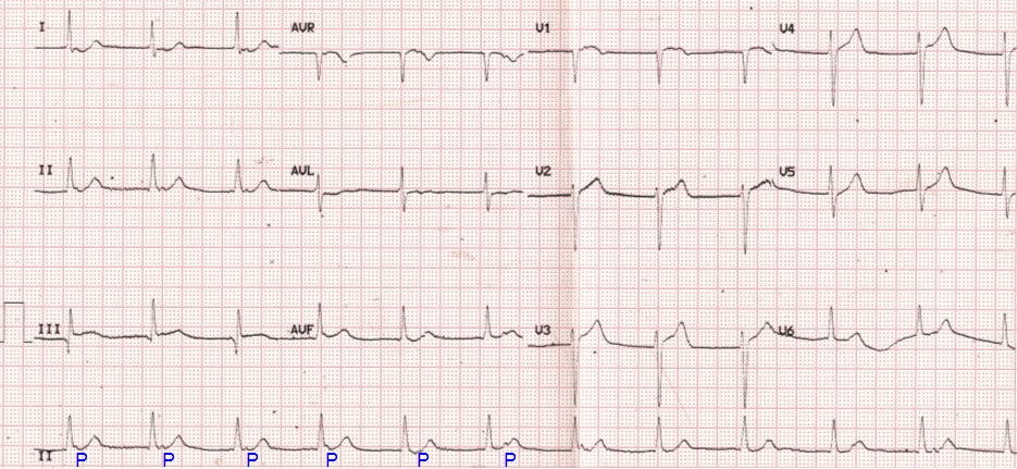
Isorhythmic AV dissociation is one of the three forms of AV (atrioventricular) dissociations which can be noted on the ECG. Complete heart block is the commonest form of AV dissociation with which we are familiar. In complete heart block the atrial rate is more than the ventricular rate, while in isorhythmic AV dissociation, the atrial and ventricular rates are almost equal. The other form of AV dissociation is interference AV dissociation in ventricular tachycardia, in which the ventricular rate is more than the atrial rate.
Isorhythmic AV dissociation occurs when the sinus rate is slowed down and junctional rate is accelerated, so that they are almost equal. The atria are captured by the sinus impulses and ventricles by the junctional impulses.
In the ECG, P waves are clearly seen to be dissociated from the QRS complex. Atrial and ventricular rates are similar. Most of the P waves occur during the ST segment. As the P waves occur during ventricular systole, atrial contraction will produce cannon waves in the jugular venous pulse because the right atrium is contracting against a closed tricuspid valve.
Reverse pulsus paradoxus in isorhythmic AV dissociation
Another interesting clinical finding which can be seen in isorhythmic AV dissociation is reverse pulsus paradoxus [1]. In reverse pulsus paradoxus, the pulse volume decreases during expiration. Here the pulse volume increases in inspiration because of increase in sinus rate which causes AV synchrony. During expiration sinus node slows down and junctional rhythm takes over, producing AV asynchrony.
Reference
- Massumi RA, Mason DT, Vera Z, Zelis R, Otero J, Amsterdam EA. Reversed pulsus paradoxus. N Engl J Med. 1973 Dec 13;289(24):1272-5.
Related Posts

About The Author
Johnson Francis
Former Professor of Cardiology, Calicut Govt. Medical Kozhikode, Kerala, India. Editor-in-Chief, BMH Medical Journal