Left ventricular hypertrophy with dysfunction – M-Mode echocardiogram
Left ventricular hypertrophy with dysfunction – M-Mode echocardiogram
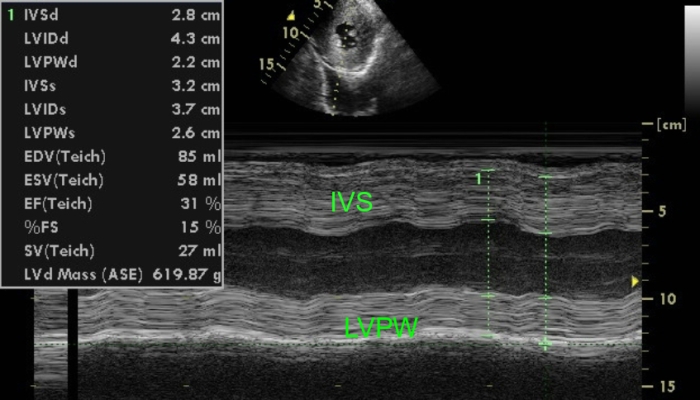
M-mode echocardiogram showing gross left ventricular hypertrophy and moderate to severe left ventricular dysfunction. Inset shows short axis view of left ventricle demonstrating severe concentric left ventricular hypertrophy. IVS: interventricular septum. LVPW: left ventricular posterior wall. IVSd: interventricular septum in diastole; LVIDd: left ventricular diastolic dimension in diastole; LVPWD: left ventricular posterior wall in diastole; IVSs: interventricular septum in systole; LVIDs: left ventricular internal dimension in systole
Left ventricular systolic dysfunction occurs in late stage of hypertrophic cardiomyopathy. In the initial stages there is only left ventricular diastolic dysfunction due to the grossly thickened ventricular myocardium with poor diastolic relaxation. In late stages, chronic overload can cause failure of the left ventricular myocardium with systolic dysfunction. In this stage there can be progressive dilatation of the left ventricular cavity.
A similar echocardiographic pattern of severe left ventricular hypertrophy with left ventricular systolic dysfunction can also occur when severe aortic stenosis goes in for left ventricular systolic dysfunction and heart failure. Same can occur in coarctation of aorta with heart failure and severe systemic hypertension with left ventricular systolic dysfunction. Thickened left ventricular myocardium with systolic dysfunction can also occur in cardiac amyloidosis.
Related Posts

About The Author
Johnson Francis
Former Professor of Cardiology, Calicut Govt. Medical Kozhikode, Kerala, India. Editor-in-Chief, BMH Medical Journal