


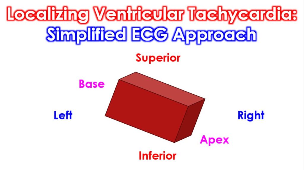

Localizing the origin of Ventricular Tachycardia from a 12-lead ECG is a critical skill for planning ablation and identifying the underlying pathology. While many algorithms exist, a stepwise simplified approach focusing on the “Four Directions” of the heart is often the most practical. To find where the VT is coming from, think of the heart as a 3D box. You use different ECG leads to check Left vs. Right, Superior vs. Inferior, and Base vs. Apex.
Step 1: Left vs. Right (Lead V1)
This is based on the Bundle Branch Block (BBB) morphology.
- LBBB Pattern (Negative V1): Origin is likely the Right Ventricle or the Interventricular Septum.
- RBBB Pattern (Positive V1): Origin is likely the Left Ventricle.
Step 2: Superior vs. Inferior (Frontal Axis – Leads II, III, aVF)
- Inferior Axis (Positive in II, III, aVF): The impulse travels downward, so the origin is Superior (e.g., Outflow Tracts).
- Superior Axis (Negative in II, III, aVF): The impulse travels upward, so the origin is Inferior (e.g., the Diaphragmatic wall).
Step 3: Base vs. Apex (Precordial Transition)
Look at where the QRS flips from mostly negative to mostly positive (the transition lead).
- Early Transition (V1–V2): Origin is Basal (posterior part of the heart).
- Late Transition (V4–V6): Origin is Apical (near the tip of the heart).
Step 4: Septum vs. Free Wall (QRS Width & Lead I)
- Septal Origin: Often has a narrower QRS because it accesses the conduction system faster.
- Free Wall Origin: Often has a wider QRS (>160ms) and specific polarity in Lead I (Positive I = Septal; Negative I = Lateral/Free Wall).