LV – RA shunt
LV – RA Shunt in perimembranous VSD
Echocardiogram video with narration
A shunt from the left ventricle to the right atrium can occur in three ways: (1) Defect in the atrioventricular septum between the septal attachments of the mitral and tricuspid valves (2) A perimembranous ventricular septal defect (VSD) with associated fenestration of the septal tricuspid leaflet so that the VSD jet is partly directed from the left ventricle across the interventricular septum through the tricuspid valve into the right atrium (3) Ventricular septal defect with tricuspid regurgitation so that the blood shunted from the left ventricle is passed immediately to the right atrium to produce a step up in oximetry.
A defect in the atrioventricular septum was described by Gerbode F et al in 5 operated cases in 1958 [1]. Anatomically this defect is possible because the septal attachment of the tricuspid valve is distal to that of the mitral valve so that there is a small region of the septum which is between the left ventricle and the right atrium, known as the atrioventricular septum. Usually the defect is congenital. But cases are on record in which the septal defect was acquired due to infective endocarditis. It is mentioned that congenital variety of defect occurs inferior to the tricuspid valve while the acquired variety is superior to the valve.
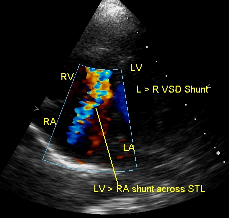
The still image shows both the jet from the left ventricle to the right ventricle across the perimembranous VSD and the jet from left ventricle to right atrium across the VSD, through the fenestration in the septal tricuspid leaflet into the right atrium.
Reference
- Gerbode F. Hultgren H. Melrose D, Osborn J. Syndrome of left ventricular-right atrial shunt: successful surgical repair of defect in five cases, with observation of bradycardia on closure. Ann Surg 1958;148(3):433-446
Related Posts


About The Author
Johnson Francis
Former Professor of Cardiology, Calicut Govt. Medical Kozhikode, Kerala, India. Editor-in-Chief, BMH Medical Journal