


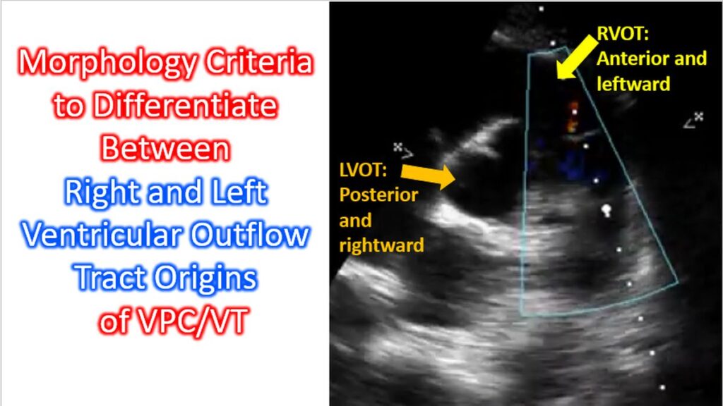
Outflow tract ventricular arrhythmias are the most common type of idiopathic ventricular arrhythmias. They typically occur in young patients. Differentiating between right ventricular outflow tract (RVOT) and left ventricular outflow tract (LVOT) origins of VPC/VT is a fundamental task in clinical electrophysiology, as it dictates the procedural approach (venous vs. arterial) for catheter ablation. Since both outflows are superiorly located, both typically present with an inferior axis (tall R-waves in II, III, and aVF). The primary differentiation relies on the horizontal plane (precordial leads). 70-80% of outflow tachycardias originate from right ventricular outflow tract. Small numbers can originate just above the outflow tracts as well, like aortic cusp ventricular tachycardia.
1. General Morphology Rules
| Feature | RVOT Origin | LVOT Origin |
|---|---|---|
| Basic Pattern | Left Bundle Branch Block (LBBB) | RBBB or Atypical LBBB |
| V1 Morphology | Deep S-wave, small/absent R-wave | Taller R-wave, smaller S-wave |
| Transition Lead | Usually V3 or later (V4, V5) | Usually V2 or earlier (V1, V2) |
| Lead I | Usually positive (leftward) | Often negative or isoelectric |
2. Specific ECG Criteria
When the transition occurs at lead V3, simple observation is often insufficient, and specific indices are used to refine the diagnosis:
- V2 Transition Ratio: This is a highly reliable metric. It is calculated by comparing the R-wave proportion during the arrhythmia (PVC) to the R-wave proportion during sinus rhythm (SR).
- V2 Transition Ratio = R/(R+S) of PVC divided by R/(R+S) of SR
- ≥ 0.6: Strongly suggests an LVOT origin.
- < 0.6: Suggests an RVOT origin.
- V2 Transition Ratio = R/(R+S) of PVC divided by R/(R+S) of SR
- V2S/V3R Ratio: Compares the S-wave in V2 to the R-wave in V3.
- A ratio ≤ 1.5 is indicative of an LVOT origin.
- R-wave Duration Index: Measured in lead V1 or V2. It is the (R-wave duration / QRS duration).
- ≥ 50%: Suggests LVOT (specifically Aortic Sinus Cusp).
- R/S Amplitude Index: Measured in lead V1 or V2.
- ≥ 30%: Suggests LVOT.
Please note that these are only generalizations based on some studies. Actually there are several sites of origin within RVOT and LVOT, with different morphologies of ECG.
3. Anatomical Correlation
The RVOT is located anterior and leftward relative to the LVOT.
- Anterior structures (RVOT) produce a late transition because the depolarization vector moves away from the anterior leads (V1-V2) initially.
- Posterior structures (LVOT) produce an earlier transition (taller R-waves in V1-V2) because the vector moves toward the anterior chest leads from the start.
4. Site-Specific Nuances
- RVOT Septum vs. Free Wall: Septal origins have a narrower QRS and earlier transition compared to free-wall origins.
- Aortic Sinus Cusps (LVOT):
- Left Coronary Cusp (LCC): Often shows a “W” or notched pattern in lead V1.
- Right Coronary Cusp (RCC): Can mimic RVOT septum but usually has a taller R-wave in V1.
Please note that the intimate and complex anatomy of the outflow tracts limits predictive value ECG criteria alone for localization for these arrhythmias.