


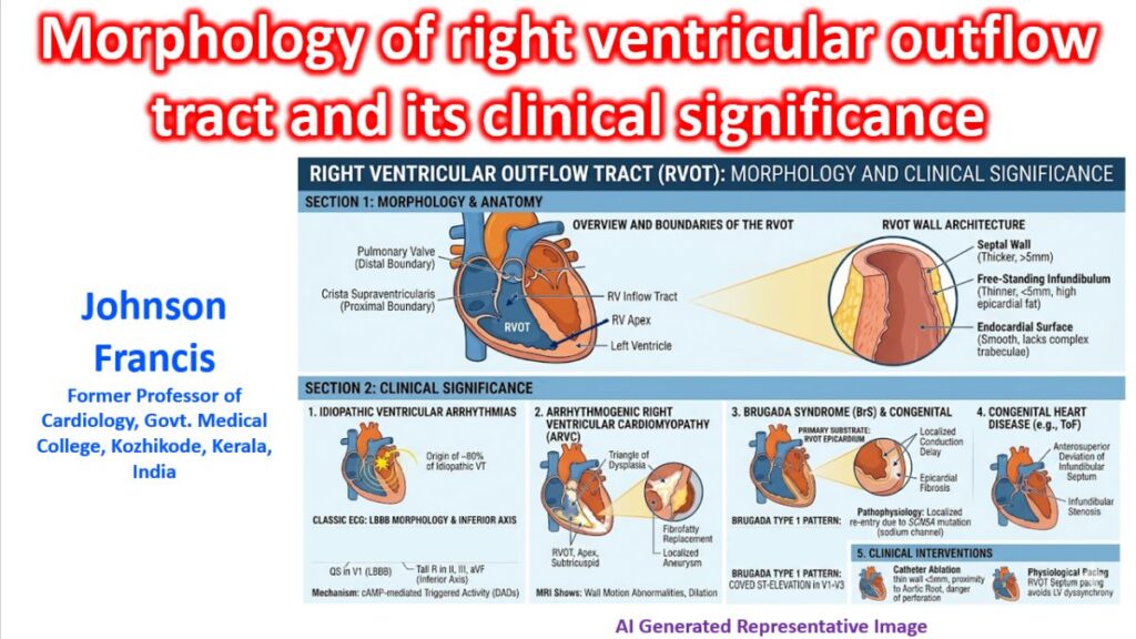

The right ventricular outflow tract (RVOT), also known as the infundibulum or conus arteriosus, is a distinct anatomical and functional entity of the right ventricle. It acts as the conduit between the trabeculated apical portion of the right ventricle and the pulmonary artery, but its unique structural characteristics make it a primary substrate for several significant cardiovascular pathologies.
Morphology and Anatomy
The RVOT is a truncated, cone-shaped, or funnel-like structure located anteriorly and to the left of the aortic root. It differs from the inflow tract of the right ventricle in both macroscopic appearance and microscopic architecture.
- Boundaries: Its proximal border is defined by the crista supraventricularis (supraventricular crest), a thick muscular ridge that separates the outflow tract from the tricuspid valve and the rough, trabeculated inflow tract. Its distal border is the basal ring of the pulmonary valve.
- Structural Components: The RVOT is composed of two main sections:
- The Septal Component: The posterior and rightward aspect, which essentially forms the superior portion of the interventricular septum.
- The Free-Standing Infundibulum: The thin-walled, anterior, and leftward free wall.
- Myoarchitecture: Unlike the main body of the right ventricle, which has a complex, intersecting meshwork of muscle fibers, the myocardial fibers in the RVOT run longitudinally and parallel to one another from the epicardium to the endocardium.
- Surface: The endocardial surface is relatively smooth, lacking the dense, coarse trabeculae carneae seen in the apical right ventricle. The epicardial surface frequently contains variable amounts of adipose tissue.
Clinical and Electrophysiological Significance
The RVOT is the most arrhythmogenic region of the structurally normal heart. Its embryological origins (derived from the embryonic bulbus cordis), thinner walls, and distinct electrophysiological properties make it highly vulnerable to both functional and structural disease states.
1. Idiopathic Ventricular Arrhythmias
The RVOT is the origin site for roughly 80% of all idiopathic ventricular tachycardias (VTs) and premature ventricular contractions (PVCs) in patients without underlying structural heart disease.
- Mechanism: These arrhythmias are typically driven by cAMP-mediated delayed afterdepolarizations (DADs) causing triggered activity. They are often provoked by exercise or catecholamine stress and are classically sensitive to adenosine.
- ECG Signature: Because the impulse originates superiorly, anteriorly, and from the right ventricle, an RVOT-VT produces a characteristic Left Bundle Branch Block (LBBB) morphology with an inferior axis (tall, positive R waves in leads II, III, and aVF).
2. Arrhythmogenic Right Ventricular Cardiomyopathy (ARVC)
The RVOT is one of the three classic points in the “triangle of dysplasia” (along with the RV apex and the subtricuspid region) affected by ARVC.
- The region is highly susceptible to progressive fibrofatty replacement of the myocardium. This creates a substrate for macro-reentrant ventricular tachycardias and can lead to localized wall motion abnormalities, dilation, and aneurysm formation visible on cardiac MRI or echocardiography.
3. Brugada Syndrome (BrS)
Recent structural and electrophysiological mapping studies have localized the primary arrhythmogenic substrate of Brugada Syndrome to the epicardium of the RVOT.
- Subtle structural micro-abnormalities, increased epicardial fibrosis, and localized conduction delays in the RVOT epicardium are responsible for the characteristic coved ST-segment elevation (Type 1 Brugada pattern) in the right precordial leads (V1-V3) and the increased vulnerability to ventricular fibrillation.
4. Congenital Heart Disease
The embryological development of the infundibulum is complex, making it a frequent site for congenital anomalies.
- Tetralogy of Fallot: The hallmark of this condition is the anterosuperior deviation of the infundibular septum. This displacement causes subvalvular right ventricular outflow tract obstruction (infundibular stenosis), which dictates the severity of right-to-left shunting and clinical cyanosis.
5. Implications for Invasive Interventions
- Catheter Ablation: The RVOT is a frequent target for the radiofrequency ablation of symptomatic PVCs and VTs. However, its exceptionally thin free wall (often < 5 mm) requires precise power titration to prevent cardiac perforation. Furthermore, the posterior/septal RVOT lies in close proximity to the aortic root (specifically the right and left coronary cusps) and the left anterior descending (LAD) artery, demanding meticulous electro-anatomical mapping before ablation.
- Physiological Pacing: To avoid pacing-induced cardiomyopathy often caused by traditional right ventricular apical pacing, the RVOT septum is frequently utilized as an alternative pacing site. Pacing from the RVOT septum engages the conduction system more naturally, resulting in a narrower QRS complex and better ventricular synchrony.