Multiple echo views in EMF
Multiple echo views in EMF
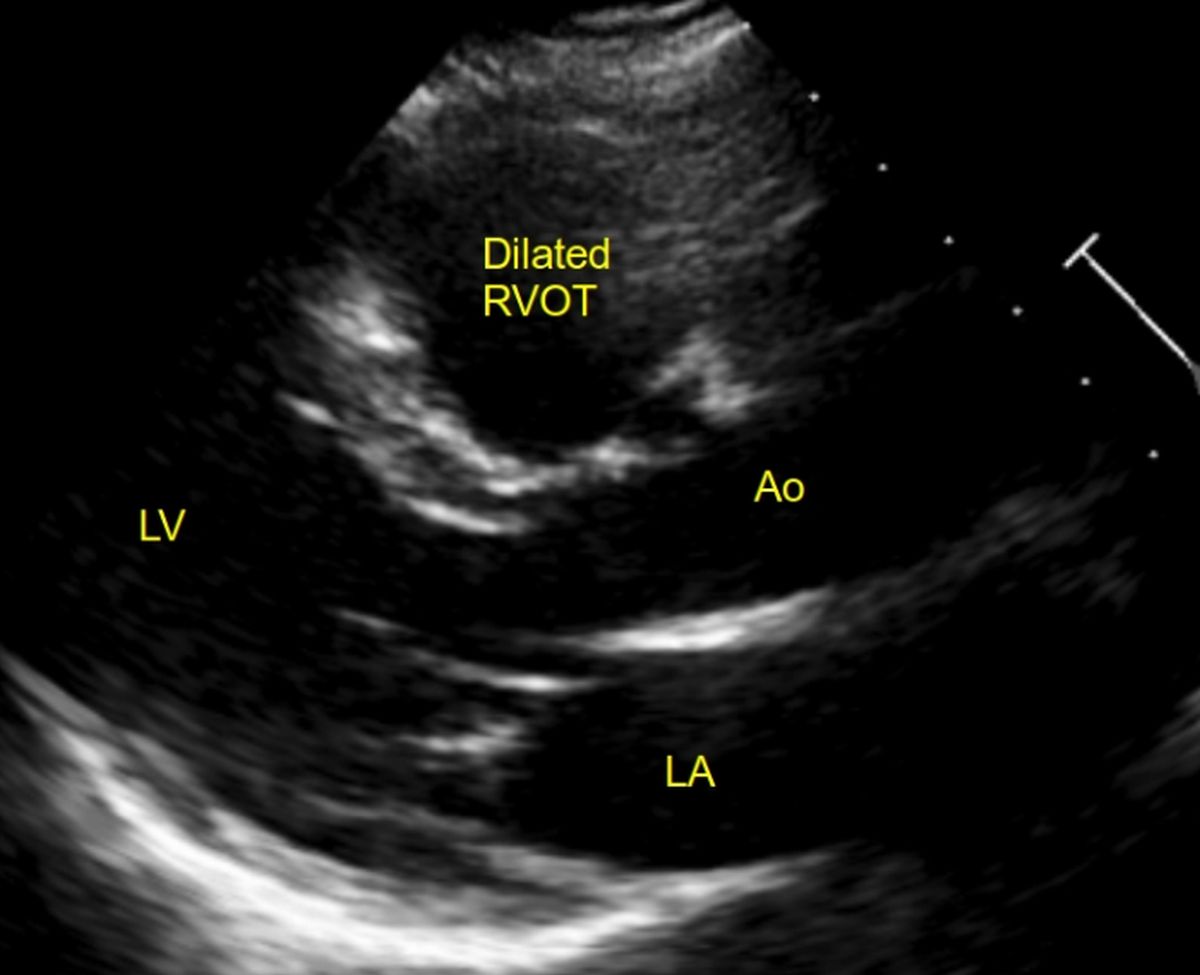
Parasternal long axis (PLAX) view shows dilated right ventricular outflow tract. This pattern alone will not give a suspicion of endomyocardial fibrosis as RVOT can be dilated along with the rest of the right ventricle in conditions causing right ventricular volume overload like an atrial septal defect as well as in Ebstein’s anomaly of the tricuspid valve. Conditions with a dilated RVOT will have a characteristic clinical finding of RVOT pulsations, along the left sternal border, in the third left intercostal space. Pulsation is not associated with a heave in conditions like Ebstein’s anomaly and right ventricular endomyocardial fibrosis, where the right ventricular pressures are not elevated.
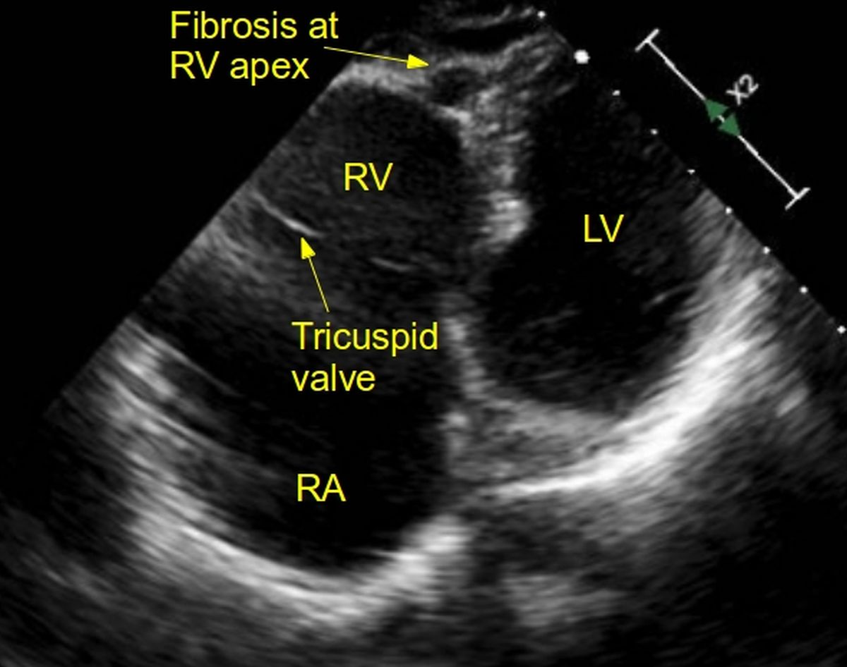
This image from a modified apical four chamber view shows the characteristic fibrosis and obliteration of right ventricular apex in EMF along with a dilated right atrium. Right ventricular cavity is not much dilated.
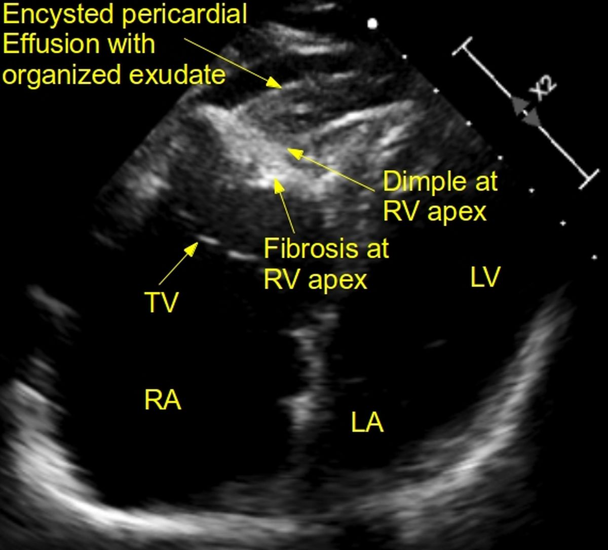
Another image from a modified apical four chamber view shows the characteristic dimple at right ventricular apex due to the fibrotic process along with a small encysted effusion nearby. The long standing exudate in the pericardial space has been organized into a clot like structure. Dilated right atrium and the fibrosis at RV apex are visible. As the prevalence of EMF is coming down drastically in our region, such images are rarely seen in an echocardiographic laboratory.
Khalil SI and colleagues have recently published data on 23 cases from Sudan, which constituted 0.5% of the 4332 echocardiograms at four hospitals [1]. Apical and ventricular wall fibrosis was noted in all cases. Other findings were atrial enlargement, AV valve regurgitation, obliteration of ventricular cavity, pericardial effusion and restrictive filling pattern. The disease involved both ventricles in 53%, right ventricle alone in 18% and left ventricle alone in 29%.
Reference
- Khalil SI, Khalil S, Tigani SE, Saad HA. Endomyocardial fibrosis in Sudan: clinical and echocardiographic features. Cardiovasc J Afr. 2017 Jul/Aug;28(4):208-214.
Related Posts


About The Author
Johnson Francis
Former Professor of Cardiology, Calicut Govt. Medical Kozhikode, Kerala, India. Editor-in-Chief, BMH Medical Journal