


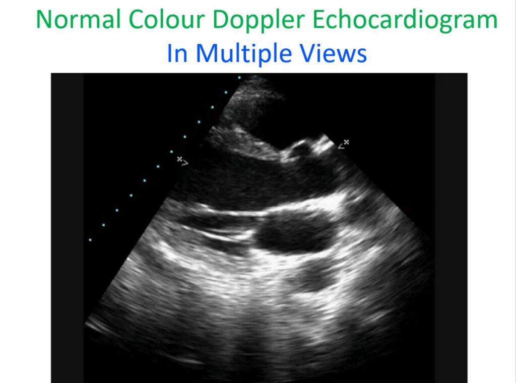

Normal Colour Doppler Echocardiogram in Multiple Views
Starting from the two dimensional parasternal long axis view, followed by colour flow mapping, M-Mode at chordal level, parasternal short axis view at aorta-left atrium level and mitral valve level, apical four chamber view and suprasternal view in sequence. Annotation is given in still pictures inserted in between the video clips.