Old inferior wall infarction and lateral ST depression
Old inferior wall infarction and lateral ST depression
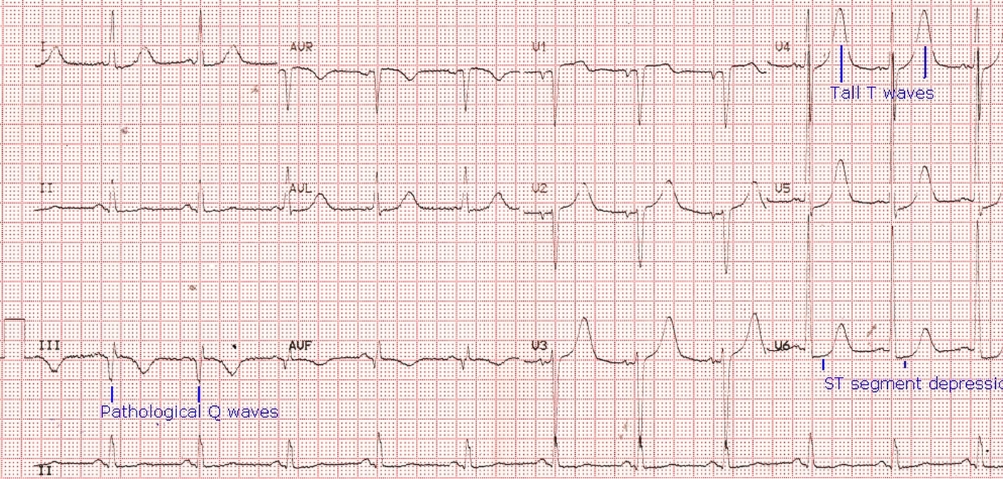
ECG shows sinus rhythm at around 75/min, with pathological Q waves in inferior leads, indicating old inferior wall myocardial infarction. Pathological Q waves are usually 40 ms in width and have an amplitude more than 25% of the ensuing R wave. T waves are inverted along with pathological Q waves in leads III and aVF. R wave voltage is high in V5 and V6 indicating left ventricular hypertrophy. The presence of mild ST segment depression in lateral leads may indicate fresh ischemia. If the ST segment depression was due to LVH, it would have been downsloping, associated with T wave inversion.
Here it is almost horizontal ST segment depression, which is usual with acute ischemia. Tall T waves in V4-V6 can occur due to any of the following: ischemia, hyperkalemia or left ventricular volume overload (diastolic overload). But tall T waves in V2 and V3 in which the predominant QRS is negative, is unlikely to be due to left ventricular volume overload, in which it is usual in leads with tall R waves. In this case it is more likely to be a manifestation of ischemia as indicated by the associated plane ST segment depression. In left ventricular volume overload, the ST segment usually shows mild upward concavity.
An interesting study correlated various ECG criteria for left ventricular hypertrophy with left ventricular mass and chamber size assessed by cardiac magnetic resonance imaging [1]. They assessed Sokolow-Lyon voltage/product, Gubner-Ungerleider voltage, Cornell voltage/product, Perugia-score and Romhilt-Estes score. They found highest predictive value with Romhilt-Estes score 4 points which had a sensitivity of 86% and specificity of 81%.
Reference
- Buchner S, Debl K, Haimerl J, Djavidani B, Poschenrieder F, Feuerbach S, Riegger GA, Luchner A. Electrocardiographic diagnosis of left ventricular hypertrophy in aortic valve disease: evaluation of ECG criteria by cardiovascular magnetic resonance. J Cardiovasc Magn Reson. 2009 Jun 1;11:18.
Related Posts


About The Author
Johnson Francis
Former Professor of Cardiology, Calicut Govt. Medical Kozhikode, Kerala, India. Editor-in-Chief, BMH Medical Journal