Papillary muscles – echocardiogram
Papillary muscles – echocardiogram
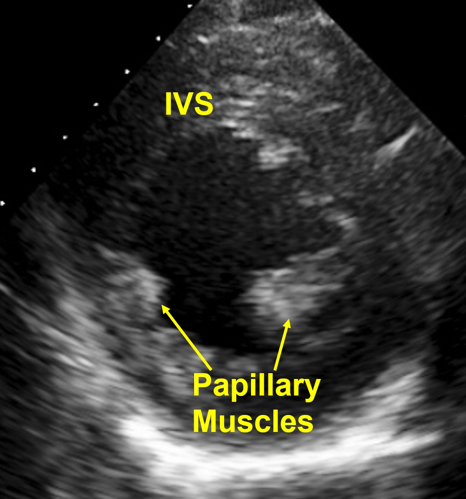
IVS: Interventricular septum
Echocardiogram from parasternal short axis view showing the two papillary muscles in the left ventricle (posteromedial and anterolateral). Papillary muscles form part of the mitral valve apparatus which prevent bulging back of the mitral leaflets during systole and thus avoiding mitral regurgitation. When the papillary muscles are ischemic and dysfunctional it results in mitral regurgitation due to papillary muscle dysfunction. If the head of the papillary muscle gets necrosed and ruptures in acute myocardial infarction, it results in acute severe mitral regurgitation. This can lead on to acute left ventricular failure which can sometimes be fatal. This is one of the mechanical complications of acute myocardial infarction which requires urgent surgical attention.
Another importance of papillary muscles is as a potential arrhythmic focus. Papillary muscle tachycardia is one of the three types of idiopathic left ventricular tachycardias, the other two being mitral annular ventricular tachycardia and fascicular ventricular tachycardia. All the three have right bundle branch block morphology as they originate in the left ventricle. All the three are ablatable ventricular tachycardias belonging to the group of monomorphic ventricular tachycardias.
About The Author
Johnson Francis
Former Professor of Cardiology, Calicut Govt. Medical Kozhikode, Kerala, India. Editor-in-Chief, BMH Medical Journal