Paradoxical motion of interventricular septum
Paradoxical motion of interventricular septum
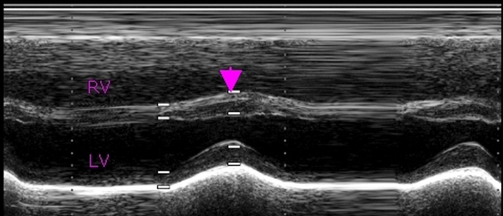
Paradoxical motion of interventricular septum: M-mode echocardiogram showing movements of the interventricular septum and posterior wall of the left ventricle (LV). Right ventricle (RV) is seen above the interventricular septum. This is an image from the parasternal view. Right ventricular free wall is adjacent the chest wall, with no echo free space in between. If there is echo free space that could be either pericardial pad of fat or pericardial effusion.
In systole, the posterior wall contracts and moves anteriorly. Normally the interventricular septum should contract and move towards the posterior wall in systole. Here the septum moves away from the posterior wall in systole (paradoxical septal motion – arrow). This occurs in situations of right ventricular volume overload like atrial septal defect and severe tricuspid regurgitation.
Interventricular septum is a common wall shared by the left ventricle and right ventricle. Normally it contracts with the left ventricle, that is why when it does not contract in synchrony with the left ventricle it is known as paradoxical septal motion. The paradoxical motion is due to change in the shape of the left ventricle. Normally the left ventricle is circular in cross section in both systole and diastole. In right ventricular volume overload, septum becomes flat or even concave towards the right ventricle in diastole. This curvature reverts in systole, producing the paradoxical motion [1]. The authors concluded that it is the diastolic change in the shape of left ventricle due to right ventricular volume overload (volume overload is also called diastolic overload) is the reason for paradoxical septal motion.
In this M-Mode echocardiogram, the vertical axis is the distance from the transducer and the horizontal axis is time. That is why it was called time-motion or TM mode earlier, now abbreviated to M-Mode.
Reference
- Weyman AE, Wann S, Feigenbaum H, Dillon JC. Mechanism of abnormal septal motion in patients with right ventricular volume overload: a cross-sectional echocardiographic study. Circulation. 1976 Aug;54(2):179-86.
Related Posts


About The Author
Johnson Francis
Former Professor of Cardiology, Calicut Govt. Medical Kozhikode, Kerala, India. Editor-in-Chief, BMH Medical Journal