


Pediatric echocardiography is the gold standard for diagnosing Congenital Heart Disease (CHD). While the field is vast, three lesions every clinician—from neonatologists to family practitioners—should be able to recognize on an echo report or clinical presentation.
1. Ventricular Septal Defect (VSD)
The Ventricular Septal Defect is the most common congenital heart lesion, accounting for nearly 30% of all CHD. It consists of a hole in the septum separating the left and right ventricles.
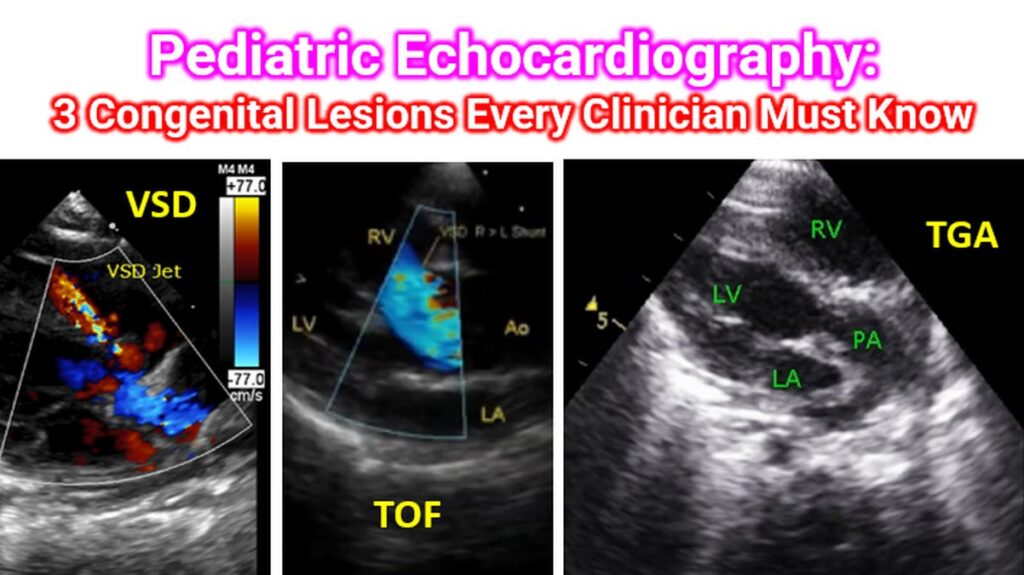
- Echo Hallmark: A high-velocity jet of blood crossing from the left ventricle (high pressure) to the right ventricle (low pressure) during systole.
- Clinician Note: Look for left-heart enlargement (LA and LV dilation) on the report. This indicates “volume overload” because the extra blood sent to the lungs eventually returns to the left side of the heart.
- Clinical Pearl: Small VSDs often produce a louder murmur (harsh, holosystolic) than large ones because the high pressure forced through a tiny hole creates more turbulence.
2. Tetralogy of Fallot (TOF)
Tetralogy of Fallot is the most common cyanotic (“blue baby”) heart defect. It is a “conotruncal” anomaly, meaning it stems from a single developmental error in the heart’s outflow tract.
It is defined by four classic features (often remembered by the acronym PROVe):
- Pulmonary Stenosis (Right Ventricular Outflow Tract Obstruction)
- Right Ventricular Hypertrophy
- Overriding Aorta (The aorta sits over the VSD instead of the left ventricle)
- Ventricular Septal Defect
- Echo Hallmark: A large “malalignment” VSD with the aortic root “straddling” or overriding the septal crest.
- Clinician Note: The severity of the child’s cyanosis is dictated by the degree of Pulmonary Stenosis. The tighter the exit to the lungs, the more deoxygenated blood is forced through the VSD into the aorta.
3. Transposition of the Great Arteries (d-TGA)
d-Transposition of the Great Arteries is a true cardiac emergency and the most common cyanotic lesion presenting in the first week of life. In TGA, the “plumbing” is switched: the aorta arises from the right ventricle, and the pulmonary artery arises from the left ventricle.
- The Result: Two separate, parallel circuits. Oxygenated blood just goes back to the lungs; deoxygenated blood just keeps circulating through the body. The baby survives only if there is a “mixing” site like a Patent Ductus Arteriosus (PDA) or Atrial Septal Defect (ASD).
- Echo Hallmark: The “Egg on a Side” appearance on X-ray, but on echo, the classic sign is seeing the two great vessels running parallel to each other rather than crossing in their normal “X” configuration. The image shows PA from LV, recognized by its bifurcation.
- Clinician Note: If you suspect TGA in a neonate, the immediate priority is keeping the ductus arteriosus open with Prostaglandin (PGE1) until surgery can be performed.
Summary for the Clinician
| Lesion | Cyanotic? | Key Echo Finding | Clinical Priority |
| VSD | No | Left-to-right systolic jet | Monitor for heart failure/weight gain |
| TOF | Yes | Overriding aorta + RVOT obstruction | Manage “Tet Spells” (knees-to-chest) |
| d-TGA | Yes (Severe) | Parallel great vessels | Maintain ductal patency (Prostaglandins) |