


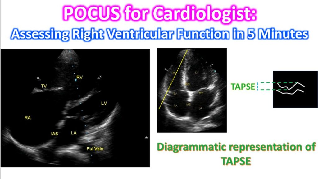

In a fast-paced clinical or emergency setting, a focused 5-minute Point-of-Care Ultrasound (POCUS) assessment of the Right Ventricle (RV) is invaluable. Because the RV is highly sensitive to afterload and preload and has a complex, crescentic geometry, a multi-parametric approach is necessary.
Here is a streamlined protocol to assess RV function efficiently.
1. Visual Assessment & Dimensions
Start with the Apical 4-Chamber (A4C) view to gauge the “big picture.”
- RV/LV Ratio: In a normal heart, the RV should be about 2/3 the size of the LV.
- Mild Dilation: RV is larger than 2/3 but smaller than the LV.
- Severe Dilation: RV is larger than the LV or forms the apex of the heart.
- McConnell’s Sign: Look for akinesia of the RV free wall with sparing of the apex—a classic (though not pathognomonic) sign of acute Pulmonary Embolism.
2. Longitudinal Function (TAPSE)
Longitudinal contraction accounts for the majority of RV stroke volume.
- Measurement: Use M-Mode in the A4C view. Place the cursor through the lateral tricuspid annulus.
- Normal Threshold: >17 mm.
- Clinical Pearl: TAPSE is highly angle-dependent. Ensure your cursor is parallel to the RV free wall motion.
3. Radial Function (FAC)
Fractional Area Change (FAC) provides a global estimate of RV systolic function by comparing end-diastolic and end-systolic areas.
FAC = {[RV Area (Diastole) – RV Area (Systole)] / RV Area (Diastole)} x 100
- Normal Threshold: >35%.
- Efficiency Tip: If the RV endocardium is difficult to trace, focus on visual “eyeballing” of inward wall motion in the Parasternal Short Axis (PSAX).
4. Pressure Overload (The “D-Sign”)
Switch to the Parasternal Short Axis (PSAX) view at the level of the papillary muscles.
- Septal Flattening: In states of RV pressure or volume overload, the interventricular septum flattens, turning the LV from an “O” shape into a “D” shape.
- Diastolic Flattening: Suggests volume overload.
- Systolic Flattening: Suggests pressure overload (e.g., Pulmonary Hypertension).
5. Right Atrial Pressure (IVC)
The Subcostal Long Axis view of the Inferior Vena Cava (IVC) serves as a proxy for Right Atrial Pressure (RAP).
| IVC Diameter | Collapsibility (Sniff Test) | Estimated RAP |
| ≤ 2.1 cm | >50% | 3 mmHg (Normal) |
| > 2.1 cm | <50% | 15 mmHg (High) |
💡 Quick Summary for the 5-Minute Scan:
- A4C: Is the RV bigger than the LV? (Dilation)
- M-Mode: Is TAPSE <17 mm? (Systolic Dysfunction)
- PSAX: Is there a “D-sign”? (Pressure/Volume Overload)
- Subcostal: Is the IVC plethoric? (High RAP/Congestion)