Prominent upper lobe vessels on CXR
Prominent upper lobe vessels on Chest X-ray
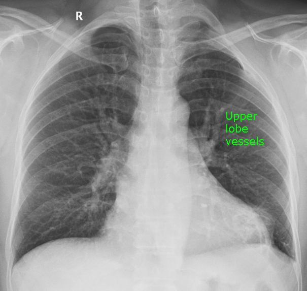
Prominent upper lobe vascular markings on X-ray chest (CXR) PA view suggests pulmonary venous hypertension. Actually they are dilated pulmonary veins, though it is not easy to differentiate between veins and arteries on CXR, hence the term prominent upper lobe vessels.
Named signs for prominent upper lobe vessels
Here are the named signs indicating prominent upper lobe vascular markings on chest X-ray:
- Antler sign [1]
- Inverted moustache sign
- Cephalization
- Redistribution
Antler sign and inverted moustache sign are self explanatory as the appearance of the prominent upper lobe vessels seen together on both sides resemble them.
Cephalization suggests the prominence of upper lobe vessels more than the lower zone vessels, which the inverse of normal.
Redistribution (upper lobe diversion, upper zone redistribution) also indicates the redistribution of pulmonary flow to the upper lobes than the lower lobes, different from the normal situation.
Lower lobe vessels are more prominent than upper lobe vessels on normal chest X-ray due to the effect of gravity. In pulmonary venous hypertension, they become less prominent due to vasoconstriction in the lower zones so that there is a redistribution of pulmonary blood flow to the upper zones.
Pulmonary venous hypertension is one of the important features of mitral stenosis which can be recognized on CXR. It can also occur in other conditions which cause elevation of left atrial pressure, which is directly transmitted to the pulmonary veins. Cephalization of pulmonary vascular marking can be a feature in heart failure.
Antler has also been described in torsion of the lung, a rare condition [2]. Torsion of the lung can be either of the whole lung or a single lobe. It can occur spontaneously in association with other anomalies. Torsion of the lung may also occur after a traumatic pneumothorax or after thoracic surgery.
References
- Han J, Xiang H, Ridley WE, Ridley LJ. Antler sign: Pulmonary venous hypertension. J Med Imaging Radiat Oncol. 2018 Oct;62 Suppl 1:13. A chest X-ray with overlay of the picture of an antler is available at the journal site.
- Hammer MM, Madan R. Clinical and imaging features in lung torsion and description of a novel imaging sign. Emerg Radiol. 2018 Apr;25(2):121-127.
Related Posts

About The Author
Johnson Francis
Former Professor of Cardiology, Calicut Govt. Medical Kozhikode, Kerala, India. Editor-in-Chief, BMH Medical Journal