Severe tricuspid regurgitation – echocardiogram
Severe tricuspid regurgitation – echocardiogram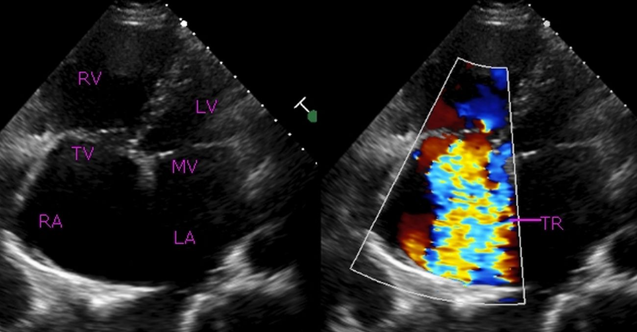
Echocardiogram in apical four chamber view shows severe tricuspid regurgitation as a large mosaic jet filling more than half of a dilated right atrium. Right ventricle is also dilated. Tricuspid valve and mitral valve are in the closed position. A large part of the interatrial septum is missing, though it is not easy to comment on interatrial septum in this view as the septum is parallel to the ultrasound beam. An echo dropout in the interatrial septum is quite common in the apical four chamber view especially at the site of foramen ovale where the septum is thin.
Trans catheter repair of tricuspid valve in high surgical risk candidates are being reported [1]. Earlier repair of tricuspid valve was using a Carpentier annuloplasty ring. This used to be done mostly during open heart surgery for repair or replacement of left sided heart valves, usually mitral and seldom as a stand alone procedure. Most cases of severe tricuspid regurgitation needing repair are secondary to mitral valve disease and severe pulmonary hypertension and hence this approach was fairly frequent.
Tricuspid annuloplasty can also be done without using a ring, known as De Vega procedure. Tang GH et al in a comparative study reported that using an annuloplasty ring is associated with improved survival and event-free survival [2].
References
- Campelo-Parada F, Perlman G, Philippon F, Ye J, Thompson C, Bédard E, Abdul-Jawad Altisent O, Del Trigo M, Leipsic J, Blanke P, Dvir D, Puri R, Webb JG, Rodés-Cabau J. First-in-Man Experience of a Novel Transcatheter Repair System for Treating Severe Tricuspid Regurgitation. J Am Coll Cardiol. J Am Coll Cardiol. 2015 Dec 8;66(22):2475-83.
- Tang GH, David TE, Singh SK, Maganti MD, Armstrong S, Borger MA. Tricuspid valve repair with an annuloplasty ring results in improved long-term outcomes. Circulation. 2006 Jul 4;114(1 Suppl):I577-81.
Related Posts


About The Author
Johnson Francis
Former Professor of Cardiology, Calicut Govt. Medical Kozhikode, Kerala, India. Editor-in-Chief, BMH Medical Journal