Straightening of left border in mitral stenosis
Straightening of left border in mitral stenosis, on X-ray chest PA view
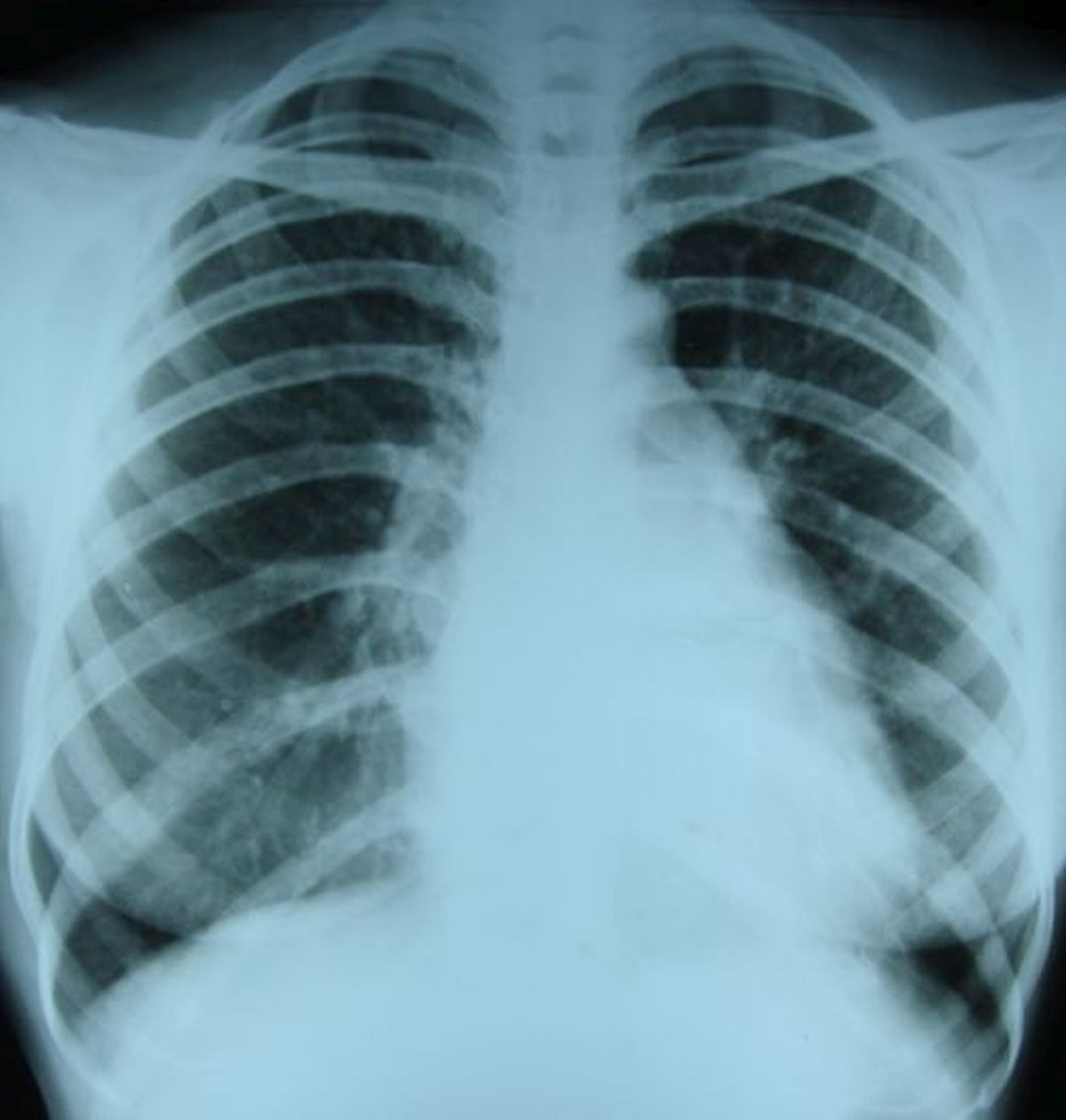 The uppermost portion on the left cardiac border is the aortic knuckle. The next slight bulge is the main pulmonary artery and the left atrial appendage is seen below that. The latter two regions are usually concave and the obliteration of the concavity contributes to the so called straightening of left border. Ao: aorta; MPA: main pulmonary artery; LAA: left atrial appendage

Straightening of the left heart border in severe mitral stenosis has been reported by Chadha et al [1]. They noted that the aortic knuckle, dilated pulmonary artery, prominent left atrial appendage and left ventricular left border are almost in a straight line. This is straightening of left border which has also been called ‘mitralization of the heart’. Their patient had presented to emergency room with features of pulmonary edema. So the X-ray showed in addition, features suggestive of bilateral pulmonary edema.
The prominent left atrial appendage is also called the ‘third mogul‘ sign. First mogul being aortic knuckle and second mogul the prominent main pulmonary artery. The bulge of the left atrial appendage will not be seen when there is mitral re-stenosis after closed mitral valvotomy, now an obsolete procedure which has given way to balloon mitral valvotomy. This is because the left atrial appendage is removed during closed mitral valvotomy as it is a common location for thrombus formation and embolization.
In another case reported by Kapoor et al, gross biatrial enlargement caused major portion of the cardiac shadow to be on the right side of the thorax, mimicking dextrocardia [2]. The left heart border was straight.
References
- S Chadha, V Shetty, A Sadiq, G Hollander, J Shani. QJM. 2013 Aug;106(8):775-6.
- Mukul C Kapoor, Anju Sarupria, Kushant Gupta, Arkalgud Sampath Kumar. Cardiomegaly Due to Left Atrial Enlargement Mimicking Dextrocardia in Chest Radiograph. Ann Card Anaesth. Oct-Dec 2013;16(4):279-80.
Related Posts


About The Author
Johnson Francis
Former Professor of Cardiology, Calicut Govt. Medical Kozhikode, Kerala, India. Editor-in-Chief, BMH Medical Journal