Systolic and Diastolic Frames From Apical Four Chamber View
Systolic and diastolic frames from apical 4C view
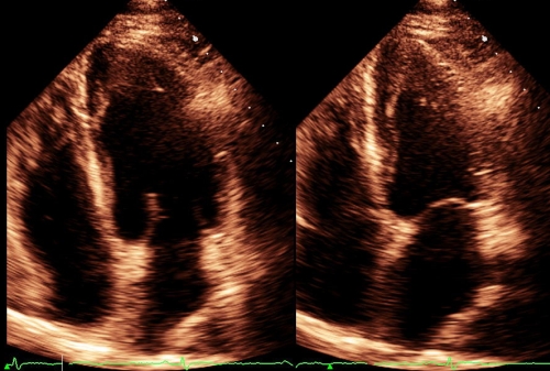
Systolic and diastolic frames from apical four chamber view on echocardiography seen on the same screen, showing all four cardiac chamers. The left image is in diastole as the mitral valve is seen to be open. Only anterior mitral leaflet is visible as jutting into the left ventricle. In the right image, the mitral valve is seen in closed position, separating the left ventricle above and the left atrium below. Part of the septal tricuspid leaflet is also seen in the closed position. See that there is a short separation between the septal attachments of the mitral and tricuspid leaflets. This region is known as the atrioventricular septum.
In apical four chamber view, chambers appear upside down as the imaging if from below upwards. Atrioventricular septum is absent in endocardial cushion defects and the two septaly attached leaflets will be at the same level. A defect in the atrioventricular septum is called as a Gerbode ventricular septal defect (Gerbode VSD). In Ebstein’s anomaly the separation between the mitral and tricuspid attachments are increased so that the septal tricuspid leaflet is displaced distally. The green tracing at the bottom of the image is the ECG for identifying the frames as systolic and diastolic, when there is a doubt. Usually systolic and diastolic frames are captured for measuring the ventricular volume and ejection fraction by the area length/Simpson’s method.
Related Posts

About The Author
Johnson Francis
Former Professor of Cardiology, Calicut Govt. Medical Kozhikode, Kerala, India. Editor-in-Chief, BMH Medical Journal