


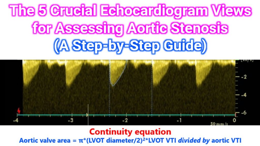

Assessing the severity of Aortic Stenosis (AS) requires a meticulous, multi-window approach. Because ultrasound is angle-dependent, failing to use multiple views can lead to an underestimation of the jet velocity – and therefore the severity – in up to 20% of patients.
The following five views are the gold standard for a comprehensive evaluation, forming the basis of the Continuity Equation used to calculate the Aortic Valve Area (AVA).
1. Parasternal Long Axis (PLAX) View
The PLAX view is the starting point for anatomic assessment and the first step in quantification.
- What to look for: Evaluate valve morphology (calcification, leafet thickening) and restricted motion (doming).
- Crucial Step: Measure the LVOT Diameter. This is done in mid-systole, from the inner-to-inner edge of the septal endocardium to the anterior mitral leaflet, usually 0.5–1.0 cm proximal to the valve.
Pro Tip: A small error in this diameter is squared in the area formula, so take multiple measurements to ensure accuracy. Some authors have recommended measurement at the annulus rather than 0.5-1.0 cm proximal to the valve.
2. Parasternal Short Axis (PSAX) View
Often called the “Mercedes-Benz” view, this provides a cross-sectional look at the valve. The name comes from the appearance of the closed aortic valve in cross section, though it is upside down!
- What to look for: This is the best view to identify the number of cusps (e.g., bicuspid vs. tricuspid).
- Crucial Step: In some patients with excellent image quality, you can perform 2D Planimetry to manually trace the valve orifice area, though Doppler methods remain the primary standard.
3. Apical 5-Chamber (A5C) View
This is the primary view for hemodynamic assessment because the ultrasound beam is usually most parallel to the flow of blood leaving the heart.
- What to look for: Use Pulsed Wave (PW) Doppler to get the LVOT VTI (Velocity Time Integral) and Continuous Wave (CW) Doppler to get the Aortic Valve VTI. DVI (Doppler Velocity Index) is a crucial dimensionless ratio between the two values.
- Crucial Step: Ensure the cursor is perfectly parallel to the jet. Use Color Doppler first to identify the “aliasing” (turbulent) jet to guide your CW placement.
4. Apical 3-Chamber (A3C) View
Also known as the Apical Long Axis view, this serves as an alternative or confirmatory window to the A5C.
- What to look for: It provides a different angle on the LVOT and aortic valve.
- Crucial Step: If the A5C yields a lower velocity than expected, the A3C may offer a better alignment with the eccentric jets often seen in calcified valves.
5. Suprasternal Notch (SSN) View
The SSN view is frequently overlooked but is vital for “picking up” the highest possible velocity.
- What to look for: This view looks “down” into the ascending aorta.
- Crucial Step: In many patients, particularly those with a dilated aorta or an eccentric jet, the highest velocity is found here rather than the apical windows. Using a PEDOF (blind) probe in this position is highly recommended for accurate grading.
PEDOF is an acronym derived from the term “Pulsed Echo DOppler Flowmeter” by the Norwegian engineers who developed it! We used to call it as the non-imaging pencil probe. You can align the probe for the best Doppler signal by keeping your eyes shut and concentrating on the Doppler sound heard in the loudspeaker of the echocardiography machine.