


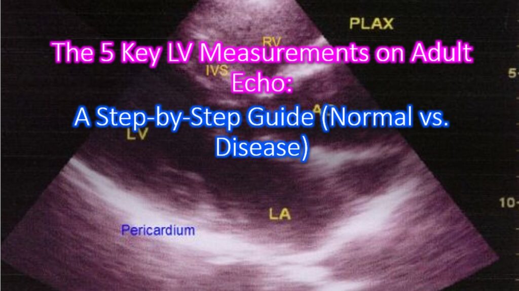

Measuring the Left Ventricle (LV) is the bread and butter of adult echocardiography. Getting these right is the difference between a routine check-up and a critical diagnosis. Here is a step-by-step breakdown of the five key measurements, how to perform them, and what those numbers actually mean. Normal values mentioned may be considered only as representative as actual value can vary with body habitus and the population under consideration.
1. LV Linear Dimensions (Internal Diameters)
This is the starting point for assessing LV size and volume. It is typically performed in the Parasternal Long Axis (PLAX) view.
- How to measure: Use 2D-guided linear measurements (M-Mode is less preferred now due to off-axis errors). Measure at the level of the mitral valve leaf tips, perpendicular to the LV long axis.
- LVIDd: LV Internal Diameter at end-diastole.
- LVIDs: LV Internal Diameter at end-systole.
- Normal vs. Disease:
- Normal: Men < 5.9 cm; Women < 5.3 cm.
- Dilated: Values above these thresholds suggest Dilated Cardiomyopathy or chronic volume overload (e.g., severe Aortic Regurgitation).
2. LV Wall Thickness (Septum and Posterior Wall)
Assessing the thickness of the walls helps differentiate between a healthy heart and one struggling against pressure.
- How to measure: Also measured in the PLAX view at end-diastole.
- IVSd: Interventricular Septum thickness.
- LVPWd: LV Posterior Wall thickness.
- Normal vs. Disease:
- Normal: ≤ 1.0 cm (Men); ≤ 0.9 cm (Women).
- Hypertrophy: > 1.2 cm is generally considered hypertrophic. This is common in Hypertension (concentric hypertrophy) or Hypertrophic Cardiomyopathy (HCM) (often asymmetric).
3. LV Ejection Fraction (LVEF)
The gold standard for “how well is the heart pumping?”
- How to measure: Use the Biplane Method of Disks (Modified Simpson’s Rule). You must trace the endocardial border in both Apical 4-Chamber and Apical 2-Chamber views at end-diastole and end-systole.
- EF = [(LVEDV – LVESV) / LVEDV] x 100
- Normal vs. Disease:
- Normal: 52% – 72% (Men); 54% – 74% (Women).
- Heart Failure (HFrEF): < 40% indicates significant systolic dysfunction.
4. Left Ventricular Mass (LVM)
While wall thickness tells you about a single “slice,” LV Mass tells you about the total weight of the muscle.
- How to measure: Usually calculated by the ultrasound software using the linear measurements (Cube formula) or 2D area-length methods. It is then indexed to body surface area (LVMI).
- Normal vs. Disease:
- Normal (Indexed): ≤ 115 g/m2 (Men); ≤ 95 g/m2 (Women).
- Elevated: High mass indicates chronic adaptation to pressure (Aortic Stenosis/Hypertension).
5. Relative Wall Thickness (RWT)
This measurement helps categorize the type of remodeling the heart is undergoing.
- How to measure: RWT = (2 x LVPWd) / LVIDd
- Normal vs. Disease:
- Normal: < 0.42.
- Concentric Hypertrophy: High RWT (> 0.42) + High LV Mass.
- Eccentric Hypertrophy: Low RWT (≤ 0.42) + High LV Mass (common in volume overload/athletes).
Pro Tip: Always ensure your image is not “foreshortened” in the apical views. If the LV looks like a football rather than a bullet, your volumes will be underestimated and your EF calculation will be wrong!